|
|
|
|
 Description
|
|
Compounds
|
||||||||||||||||||||||||||||||||||||||||||||||||||||
Chains, Units
Summary Information (see also Sequences/Alignments below) |
Ligands, Modified Residues, Ions (4, 5)| Asymmetric Unit (4, 5) Biological Unit 1 (3, 8) |
Sites (5, 5)
Asymmetric Unit (5, 5)
|
SS Bonds (6, 6)
Asymmetric Unit
|
||||||||||||||||||||||||||||
Cis Peptide Bonds (0, 0)| (no "Cis Peptide Bond" information available for 1LU0) |
SAPs(SNPs)/Variants (0, 0)| (no "SAP(SNP)/Variant" information available for 1LU0) |
PROSITE Motifs (1, 2)
Asymmetric Unit (1, 2)
|
||||||||||||||||||||||||||||||||||||||||||||||||
Exons (0, 0)| (no "Exon" information available for 1LU0) |
Sequences/Alignments
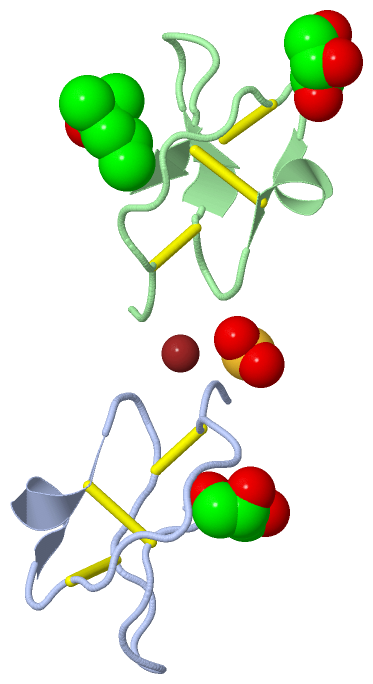
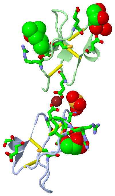
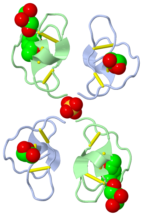
Asymmetric UnitChain A from PDB Type:PROTEIN Length:29 aligned with ITR1_CUCMA | P01074 from UniProtKB/Swiss-Prot Length:29 Alignment length:29 10 20 ITR1_CUCMA 1 RVCPRILMECKKDSDCLAECVCLEHGYCG 29 SCOP domains d1lu0a_ A: Trypsin inhibitor SCOP domains CATH domains ----------------------------- CATH domains Pfam domains ----------------------------- Pfam domains SAPs(SNPs) ----------------------------- SAPs(SNPs) PROSITE --SQUASH_INHIBITOR ------- PROSITE Transcript ----------------------------- Transcript 1lu0 A 1 RVCPRILLECKKDSDCLAECVCLEHGYCG 29 10 20 Chain B from PDB Type:PROTEIN Length:29 aligned with ITR1_CUCMA | P01074 from UniProtKB/Swiss-Prot Length:29 Alignment length:29 10 20 ITR1_CUCMA 1 RVCPRILMECKKDSDCLAECVCLEHGYCG 29 SCOP domains d1lu0b_ B: Trypsin inhibitor SCOP domains CATH domains ----------------------------- CATH domains Pfam domains (1) Squash-1lu0B01 B:1-29 Pfam domains (1) Pfam domains (2) Squash-1lu0B02 B:1-29 Pfam domains (2) SAPs(SNPs) ----------------------------- SAPs(SNPs) PROSITE --SQUASH_INHIBITOR ------- PROSITE Transcript ----------------------------- Transcript 1lu0 B 1 RVCPRILLECKKDSDCLAECVCLEHGYCG 29 10 20
|
||||||||||||||||||||
SCOP Domains (1, 2)
Asymmetric Unit
 |
CATH Domains (0, 0)| (no "CATH Domain" information available for 1LU0) |
Pfam Domains (1, 2)
Asymmetric Unit
|
Gene Ontology (6, 6)|
Asymmetric Unit(hide GO term definitions) Chain A,B (ITR1_CUCMA | P01074)
|
||||||||||||||||||||||||||||||||||||||||||||||||||||||
Interactive Views
|
|||||||||||||||||||||||||||||||||||||||||||||||||||||||||||||||||||||||||||||||||||||||||||||||||||||||||||||||||||||||||||||||||||||||||||||||||||||||||||||||||||||||||||||||||||||||||
Still Images
|
||||||||||||||||
Databases
|
||||||||||||||||||||||||||||||||||||||||||||||||||||||||||||||||||||||||||||||||||||||||||||||||||||||||||||||||||||||||||||||||||||||||||||||||||||||||||||||||
Analysis Tools
|
|||||||||||||||||||||||||||||||||||||||||||||||||||||||||||||
Entries Sharing at Least One Protein Chain (UniProt ID)
Related Entries Specified in the PDB File
|
|