|
|
|
|
 Description
|
|
Compounds
|
||||||||||||||||||||||||
Chains, Units
Summary Information (see also Sequences/Alignments below) |
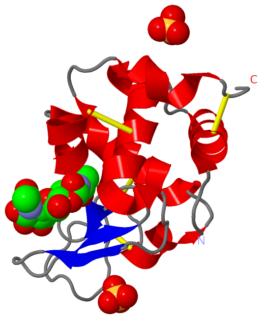
Ligands, Modified Residues, Ions (2, 3)| Asymmetric/Biological Unit (2, 3) |
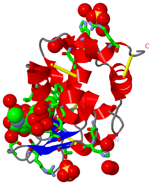
Sites (3, 3)
Asymmetric Unit (3, 3)
|
SS Bonds (4, 4)
Asymmetric/Biological Unit
|
||||||||||||||||||||
Cis Peptide Bonds (0, 0)| (no "Cis Peptide Bond" information available for 1LJN) |
SAPs(SNPs)/Variants (0, 0)| (no "SAP(SNP)/Variant" information available for 1LJN) |
PROSITE Motifs (2, 2)
Asymmetric/Biological Unit (2, 2)
|
||||||||||||||||||||||||||||||||
Exons (0, 0)| (no "Exon" information available for 1LJN) |
Sequences/Alignments
Asymmetric/Biological UnitChain A from PDB Type:PROTEIN Length:129 aligned with LYSC_MELGA | P00703 from UniProtKB/Swiss-Prot Length:147 Alignment length:129 28 38 48 58 68 78 88 98 108 118 128 138 LYSC_MELGA 19 KVYGRCELAAAMKRLGLDNYRGYSLGNWVCAAKFESNFNTHATNRNTDGSTDYGILQINSRWWCNDGRTPGSKNLCNIPCSALLSSDITASVNCAKKIASGGNGMNAWVAWRNRCKGTDVHAWIRGCRL 147 SCOP domains d1ljna_ A: Lysozyme SCOP domains CATH domains 1ljnA00 A:1-129 [code=1.10.530.10, no name defined] CATH domains Pfam domains --------------------------------------------------------------------------------------------------------------------------------- Pfam domains SAPs(SNPs) --------------------------------------------------------------------------------------------------------------------------------- SAPs(SNPs) PROSITE (1) LACTALBUMIN_LYSOZYME_2 PDB: A:1-129 UniProt: 19-147 PROSITE (1) PROSITE (2) ---------------------------------------------------------------------------LACTALBUMIN_LYSOZYM----------------------------------- PROSITE (2) Transcript --------------------------------------------------------------------------------------------------------------------------------- Transcript 1ljn A 1 KVYGRCELAAAMKRLGLDNYRGYSLGNWVCAAKFESNFNTHATNRNTDGSTDYGILQINSRWWCNDGRTPGSKNLCNIPCSALLSSDITASVNCAKKIASGGNGMNAWVAWRNRCKGTDVHAWIRGCRL 129 10 20 30 40 50 60 70 80 90 100 110 120
|
||||||||||||||||||||
SCOP Domains (1, 1)
Asymmetric/Biological Unit
 |
CATH Domains (1, 1)
Asymmetric/Biological Unit
|
Pfam Domains (0, 0)| (no "Pfam Domain" information available for 1LJN) |
Gene Ontology (12, 12)|
Asymmetric/Biological Unit(hide GO term definitions) Chain A (LYSC_MELGA | P00703)
|
||||||||||||||||||||||||||||||||||||||||||||||||||||||||||||||||||||||||||||||||||||||||||
Interactive Views
|
|||||||||||||||||||||||||||||||||||||||||||||||||||||||||||||||||||||||||||||||||||||||||||||||||||||||||||||||||||||||||||||||||||||||||||
Still Images
|
||||||||||||||||
Databases
|
||||||||||||||||||||||||||||||||||||||||||||||||||||||||||||||||||||||||||||||||||||||||||||||||||||||||||||||||||||||||||||||||||||||||||||||||||||||||||||||||
Analysis Tools
|
|||||||||||||||||||||||||||||||||||||||||||||||||||||||||||||
Entries Sharing at Least One Protein Chain (UniProt ID)
Related Entries Specified in the PDB File
|
|