Show PDB file:   
         Plain Text   HTML   (compressed file size)
QuickSearch:   
by PDB,NDB,UniProt,PROSITE Code or Search Term(s)  
(-)Asymmetric Unit
(-)Asym. Unit - sites
(-)Biological Unit 1
(-)Biol. Unit 1 - sites
(-)Biological Unit 2
collapse expand < >
Image Asymmetric Unit
Asymmetric Unit  (Jmol Viewer)
Image Asym. Unit - sites
Asym. Unit - sites  (Jmol Viewer)
Image Biological Unit 1
Biological Unit 1  (Jmol Viewer)
Image Biol. Unit 1 - sites
Biol. Unit 1 - sites  (Jmol Viewer)
Image Biological Unit 2
Biological Unit 2  (Jmol Viewer)

(-) Description

Title :  HIGH RESOLUTION LIGANDED STRUCTURE OF PHOSPHOSERINE PHOSPHATASE (PI COMPLEX)
 
Authors :  W. Wang, H. S. Cho, R. Kim, J. Jancarik, H. Yokota, H. H. Nguyen, I. V. Grigoriev, D. E. Wemmer, S. H. Kim, Berkeley Structural Genomics Center (Bsgc)
Date :  15 Mar 02  (Deposition) - 03 Apr 02  (Release) - 24 Feb 09  (Revision)
Method :  X-RAY DIFFRACTION
Resolution :  1.48
Chains :  Asym. Unit :  A,B
Biol. Unit 1:  A  (1x)
Biol. Unit 2:  B  (1x)
Keywords :  Rossmann Fold, Four-Helix Bundle, B-Hairpin, Structural Genomics, Bsgc Structure Funded By Nih, Protein Structure Initiative, Psi, Berkeley Structural Genomics Center, Hydrolase (Keyword Search: [Gene Ontology, PubMed, Web (Google))
 
Reference :  W. Wang, H. S. Cho, R. Kim, J. Jancarik, H. Yokota, H. H. Nguyen, I. V. Grigoriev, D. E. Wemmer, S. H. Kim
Structural Characterization Of The Reaction Pathway In Phosphoserine Phosphatase: Crystallographic "Snapshots" Of Intermediate States.
J. Mol. Biol. V. 319 421 2002
PubMed-ID: 12051918  |  Reference-DOI: 10.1016/S0022-2836(02)00324-8
(for further references see the PDB file header)

(-) Compounds

Molecule 1 - PHOSPHOSERINE PHOSPHATASE
    ChainsA, B
    EC Number3.1.3.3
    EngineeredYES
    Expression SystemESCHERICHIA COLI
    Expression System PlasmidPET21A
    Expression System StrainB384(DE3)/PSJS1244
    Expression System Taxid562
    Expression System Vector TypePLASMID
    GeneMJ1594
    Organism ScientificMETHANOCALDOCOCCUS JANNASCHII
    Organism Taxid2190
    SynonymPSP, O-PHOSPHOSERINE PHOSPHOHYDROLASE, PSPASE

 Structural Features

(-) Chains, Units

  12
Asymmetric Unit AB
Biological Unit 1 (1x)A 
Biological Unit 2 (1x) B

Summary Information (see also Sequences/Alignments below)

(-) Ligands, Modified Residues, Ions  (3, 10)

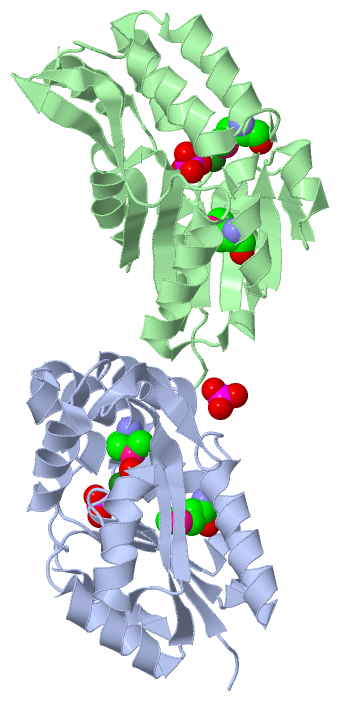
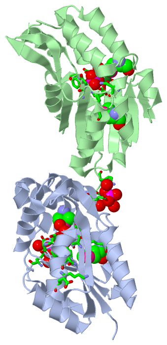
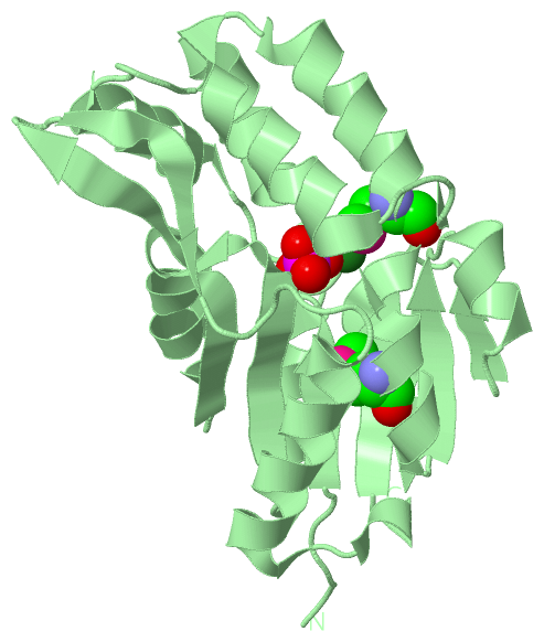
Asymmetric Unit (3, 10)
No.NameCountTypeFull Name
1MG2Ligand/IonMAGNESIUM ION
2MSE4Mod. Amino AcidSELENOMETHIONINE
3PO44Ligand/IonPHOSPHATE ION
Biological Unit 1 (2, 5)
No.NameCountTypeFull Name
1MG-1Ligand/IonMAGNESIUM ION
2MSE2Mod. Amino AcidSELENOMETHIONINE
3PO43Ligand/IonPHOSPHATE ION
Biological Unit 2 (2, 3)
No.NameCountTypeFull Name
1MG-1Ligand/IonMAGNESIUM ION
2MSE2Mod. Amino AcidSELENOMETHIONINE
3PO41Ligand/IonPHOSPHATE ION

(-) Sites  (6, 6)

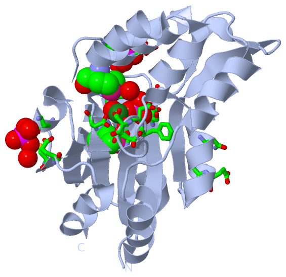
Asymmetric Unit (6, 6)
No.NameEvidenceResiduesDescription
1AC1SOFTWAREMG B:221 , ASP B:511 , PHE B:512 , ASP B:513 , SER B:599 , GLY B:600 , GLY B:601 , LYS B:644 , ASN B:670 , HOH B:857 , HOH B:960BINDING SITE FOR RESIDUE PO4 B 220
2AC2SOFTWAREPO4 B:220 , ASP B:511 , ASP B:513 , ASP B:667 , HOH B:960BINDING SITE FOR RESIDUE MG B 221
3AC3SOFTWAREASP A:11 , PHE A:12 , ASP A:13 , SER A:99 , GLY A:100 , LYS A:144 , ASN A:170 , MG A:721 , HOH A:831 , HOH A:956BINDING SITE FOR RESIDUE PO4 A 720
4AC4SOFTWAREASP A:11 , ASP A:13 , SER A:14 , ASP A:167 , PO4 A:720 , HOH A:956BINDING SITE FOR RESIDUE MG A 721
5AC5SOFTWAREASN A:48 , GLU A:50 , HOH A:815 , HOH A:832 , HOH A:939 , LYS B:504BINDING SITE FOR RESIDUE PO4 A 230
6AC6SOFTWAREASN A:107 , LYS A:110 , GLU A:111 , LYS A:191 , CYS A:197 , GLU A:199 , HOH A:870 , HOH A:1099BINDING SITE FOR RESIDUE PO4 A 240

(-) SS Bonds  (0, 0)

(no "SS Bond" information available for 1L7M)

(-) Cis Peptide Bonds  (0, 0)

(no "Cis Peptide Bond" information available for 1L7M)

 Sequence-Structure Mapping

(-) SAPs(SNPs)/Variants  (0, 0)

(no "SAP(SNP)/Variant" information available for 1L7M)

(-) PROSITE Motifs  (0, 0)

(no "PROSITE Motif" information available for 1L7M)

(-) Exons   (0, 0)

(no "Exon" information available for 1L7M)

(-) Sequences/Alignments

Asymmetric Unit
   Reformat: Number of residues per line =  ('0' or empty: single-line sequence representation)
  Number of residues per labelling interval =   
  UniProt sequence: complete  aligned part    
   Show mapping: SCOP domains CATH domains Pfam domains Secondary structure (by author)
SAPs(SNPs) PROSITE motifs Exons
(details for a mapped element are shown in a popup box when the mouse pointer rests over it)
Chain A from PDB  Type:PROTEIN  Length:209
 aligned with SERB_METJA | Q58989 from UniProtKB/Swiss-Prot  Length:211

    Alignment length:209
                                    12        22        32        42        52        62        72        82        92       102       112       122       132       142       152       162       172       182       192       202         
           SERB_METJA     3 KKKKLILFDFDSTLVNNETIDEIAREAGVEEEVKKITKEAMEGKLNFEQSLRKRVSLLKDLPIEKVEKAIKRITPTEGAEETIKELKNRGYVVAVVSGGFDIAVNKIKEKLGLDYAFANRLIVKDGKLTGDVEGEVLKENAKGEILEKIAKIEGINLEDTVAVGDGANDISMFKKAGLKIAFCAKPILKEKADICIEKRDLREILKYIK 211
               SCOP domains d1l7ma_ A: Phosphoserine phosphatase                                                                                                                                                                              SCOP domains
               CATH domains 1l7mA01       1l7mA02 A:17-77 Phosphoserine phosphatase; domain 2          1l7mA01 A:3-16,A:78-210  [code=3.40.50.1000, no name defined]                                                                        - CATH domains
               Pfam domains ----------------------------------------------------------------------------------------------------------------------------------------------------------------------------------------------------------------- Pfam domains
         Sec.struct. author ...eeeeee........hhhhhhhhhh.hhhhhhhhhhhhhh...hhhhhhhhhhhh....hhhhhhhhhhh.....hhhhhhhhhhhh.eeeeeeeeeehhhhhhhhhhhh..eeeeeeeeee..eeeeeee......hhhhhhhhhhhhhhh.hhh.eeeee.hhhhhhhhhhh.eeeee..hhhhhh...eee...hhhhhhhhh. Sec.struct. author
                 SAPs(SNPs) ----------------------------------------------------------------------------------------------------------------------------------------------------------------------------------------------------------------- SAPs(SNPs)
                    PROSITE ----------------------------------------------------------------------------------------------------------------------------------------------------------------------------------------------------------------- PROSITE
                 Transcript ----------------------------------------------------------------------------------------------------------------------------------------------------------------------------------------------------------------- Transcript
                 1l7m A   3 KKKKLILFDFDSTLVNNETIDEIAREAGVEEEVKKITKEAmEGKLNFEQSLRKRVSLLKDLPIEKVEKAIKRITPTEGAEETIKELKNRGYVVAVVSGGFDIAVNKIKEKLGLDYAFANRLIVKDGKLTGDVEGEVLKENAKGEILEKIAKIEGINLEDTVAVGDGANDISmFKKAGLKIAFCAKPILKEKADICIEKRDLREILKYIK 211
                                    12        22        32        42|       52        62        72        82        92       102       112       122       132       142       152       162       172 |     182       192       202         
                                                                   43-MSE                                                                                                                            174-MSE                                 

Chain B from PDB  Type:PROTEIN  Length:210
 aligned with SERB_METJA | Q58989 from UniProtKB/Swiss-Prot  Length:211

    Alignment length:210
                                    11        21        31        41        51        61        71        81        91       101       111       121       131       141       151       161       171       181       191       201       211
           SERB_METJA     2 EKKKKLILFDFDSTLVNNETIDEIAREAGVEEEVKKITKEAMEGKLNFEQSLRKRVSLLKDLPIEKVEKAIKRITPTEGAEETIKELKNRGYVVAVVSGGFDIAVNKIKEKLGLDYAFANRLIVKDGKLTGDVEGEVLKENAKGEILEKIAKIEGINLEDTVAVGDGANDISMFKKAGLKIAFCAKPILKEKADICIEKRDLREILKYIK 211
               SCOP domains d1l7mb_ B: Phosphoserine phosphatase                                                                                                                                                                               SCOP domains
               CATH domains 1l7mB01        1l7mB02 B:517-577 Phosphoserine phosphatase; domain 2        1l7mB01 B:502-516,B:578-710  [code=3.40.50.1000, no name defined]                                                                    - CATH domains
           Pfam domains (1) ---Hydrolase-1l7mB01 B:505-679                                                                                                                                                    -------------------------------- Pfam domains (1)
           Pfam domains (2) ---Hydrolase-1l7mB02 B:505-679                                                                                                                                                    -------------------------------- Pfam domains (2)
         Sec.struct. author .....eeee.........hhhhhhhhhh.hhhhhhhhhhhhhh...hhhhhhhhhhhh....hhhhhhhhhhh.....hhhhhhhhhhhh..eeeeeeeeehhhhhhhhhhhh..eeeeeeeeee..eeeeeee......hhhhhhhhhhhhhhh.hhh.eeeee.hhhhhhhhhhh.eeeee..hhhhhh...eee...hhhhhhhhh. Sec.struct. author
                 SAPs(SNPs) ------------------------------------------------------------------------------------------------------------------------------------------------------------------------------------------------------------------ SAPs(SNPs)
                    PROSITE ------------------------------------------------------------------------------------------------------------------------------------------------------------------------------------------------------------------ PROSITE
                 Transcript ------------------------------------------------------------------------------------------------------------------------------------------------------------------------------------------------------------------ Transcript
                 1l7m B 502 EKKKKLILFDFDSTLVNNETIDEIAREAGVEEEVKKITKEAmEGKLNFEQSLRKRVSLLKDLPIEKVEKAIKRITPTEGAEETIKELKNRGYVVAVVSGGFDIAVNKIKEKLGLDYAFANRLIVKDGKLTGDVEGEVLKENAKGEILEKIAKIEGINLEDTVAVGDGANDISmFKKAGLKIAFCAKPILKEKADICIEKRDLREILKYIK 711
                                   511       521       531       541 |     551       561       571       581       591       601       611       621       631       641       651       661       671  |    681       691       701       711
                                                                   543-MSE                                                                                                                            674-MSE                                 

   Legend:   → Mismatch (orange background)
  - → Gap (green background, '-', border residues have a numbering label)
    → Modified Residue (blue background, lower-case, 'x' indicates undefined single-letter code, labelled with number + name)
  x → Chemical Group (purple background, 'x', labelled with number + name, e.g. ACE or NH2)
  extra numbering lines below/above indicate numbering irregularities and modified residue names etc., number ends below/above '|'

 Classification and Annotation

(-) SCOP Domains  (1, 2)

Asymmetric Unit

(-) CATH Domains  (2, 4)

Asymmetric Unit
(-)
Class: Alpha Beta (26913)

(-) Pfam Domains  (1, 2)

Asymmetric Unit
(-)
Clan: HAD (186)

(-) Gene Ontology  (10, 10)

Asymmetric Unit(hide GO term definitions)
Chain A,B   (SERB_METJA | Q58989)
molecular function
    GO:0016787    hydrolase activity    Catalysis of the hydrolysis of various bonds, e.g. C-O, C-N, C-C, phosphoric anhydride bonds, etc. Hydrolase is the systematic name for any enzyme of EC class 3.
    GO:0000287    magnesium ion binding    Interacting selectively and non-covalently with magnesium (Mg) ions.
    GO:0046872    metal ion binding    Interacting selectively and non-covalently with any metal ion.
    GO:0016791    phosphatase activity    Catalysis of the hydrolysis of phosphoric monoesters, releasing inorganic phosphate.
    GO:0004647    phosphoserine phosphatase activity    Catalysis of the reaction: L(or D)-O-phosphoserine + H2O = L(or D)-serine + phosphate.
biological process
    GO:0006564    L-serine biosynthetic process    The chemical reactions and pathways resulting in the formation of L-serine, the L-enantiomer of serine, i.e. (2S)-2-amino-3-hydroxypropanoic acid.
    GO:0008652    cellular amino acid biosynthetic process    The chemical reactions and pathways resulting in the formation of amino acids, organic acids containing one or more amino substituents.
    GO:0016311    dephosphorylation    The process of removing one or more phosphoric (ester or anhydride) residues from a molecule.
    GO:0008152    metabolic process    The chemical reactions and pathways, including anabolism and catabolism, by which living organisms transform chemical substances. Metabolic processes typically transform small molecules, but also include macromolecular processes such as DNA repair and replication, and protein synthesis and degradation.
cellular component
    GO:0005737    cytoplasm    All of the contents of a cell excluding the plasma membrane and nucleus, but including other subcellular structures.

 Visualization

(-) Interactive Views

Asymmetric Unit
  Complete Structure
    Jena3D(integrated viewing of ligand, site, SAP, PROSITE, SCOP information)
    WebMol | AstexViewer[tm]@PDBe
(Java Applets, require no local installation except for Java; loading may be slow)
    STRAP
(Java WebStart application, automatic local installation, requires Java; full application with system access!)
    RasMol
(require local installation)
    Molscript (VRML)
(requires installation of a VRML viewer; select preferred view via VRML and generate a mono or stereo PDF format file)
 
  Ligands, Modified Residues, Ions
    MG  [ RasMol | Jena3D ]  +environment [ RasMol | Jena3D ]
    MSE  [ RasMol | Jena3D ]  +environment [ RasMol | Jena3D ]
    PO4  [ RasMol | Jena3D ]  +environment [ RasMol | Jena3D ]
 
  Sites
    AC1  [ RasMol ]  +environment [ RasMol ]
    AC2  [ RasMol ]  +environment [ RasMol ]
    AC3  [ RasMol ]  +environment [ RasMol ]
    AC4  [ RasMol ]  +environment [ RasMol ]
    AC5  [ RasMol ]  +environment [ RasMol ]
    AC6  [ RasMol ]  +environment [ RasMol ]
 
  Cis Peptide Bonds
(no "Cis Peptide Bonds" information available for 1l7m)
 
Biological Units
  Complete Structure
    Biological Unit 1  [ Jena3D ]
    Biological Unit 2  [ Jena3D ]

(-) Still Images

Jmol
  protein: cartoon or spacefill or dots and stick; nucleic acid: cartoon and stick; ligands: spacefill; active site: stick
Molscript
  protein, nucleic acid: cartoon; ligands: spacefill; active site: ball and stick

 Databases and Analysis Tools

(-) Databases

Access by PDB/NDB ID
  1l7m
    Family and Domain InformationProDom | SYSTERS
    General Structural InformationGlycoscienceDB | MMDB | NDB | OCA | PDB | PDBe | PDBj | PDBsum | PDBWiki | PQS | PROTEOPEDIA
    Orientation in MembranesOPM
    Protein SurfaceSURFACE
    Secondary StructureDSSP (structure derived) | HSSP (homology derived)
    Structural GenomicsGeneCensus
    Structural NeighboursCE | VAST
    Structure ClassificationCATH | Dali | SCOP
    Validation and Original DataBMRB Data View | BMRB Restraints Grid | EDS | PROCHECK | RECOORD | WHAT_CHECK
 
Access by UniProt ID/Accession number
  SERB_METJA | Q58989
    Comparative Protein Structure ModelsModBase
    Genomic InformationEnsembl
    Protein-protein InteractionDIP
    Sequence, Family and Domain InformationInterPro | Pfam | SMART | UniProtKB/SwissProt
 
Access by Enzyme Classificator   (EC Number)
  3.1.3.3
    General Enzyme InformationBRENDA | EC-PDB | Enzyme | IntEnz
    PathwayKEGG | MetaCyc
 
Access by Disease Identifier   (MIM ID)
  (no 'MIM ID' available)
    Disease InformationOMIM
 
Access by GenAge ID
  (no 'GenAge ID' available)
    Age Related InformationGenAge

(-) Analysis Tools

Access by PDB/NDB ID
    Domain InformationXDom
    Interatomic Contacts of Structural UnitsCSU
    Ligand-protein ContactsLPC
    Protein CavitiescastP
    Sequence and Secondary StructurePDBCartoon
    Structure AlignmentSTRAP(Java WebStart application, automatic local installation, requires Java; full application with system access!)
    Structure and Sequence BrowserSTING
 
Access by UniProt ID/Accession number
  SERB_METJA | Q58989
    Protein Disorder PredictionDisEMBL | FoldIndex | GLOBPLOT (for more information see DisProt)

 Related Entries

(-) Entries Sharing at Least One Protein Chain (UniProt ID)

UniProtKB/Swiss-Prot
        SERB_METJA | Q589891f5s 1j97 1l7n 1l7o 1l7p

(-) Related Entries Specified in the PDB File

1f5s CRYSTAL STRUCTURE OF PHOSPHOSERINE PHOSPHATASE FROM METHANOCOCCUS JANNASCHII
1j97 PHOSPHO-ASPARTYL INTERMEDIATE ANALOGUE OF PHOSPHOSERINE PHOSPHATASE CLASSIFICATION HYDROLASE RELATED ID: BSGCAIR30380 RELATED DB: TARGETDB