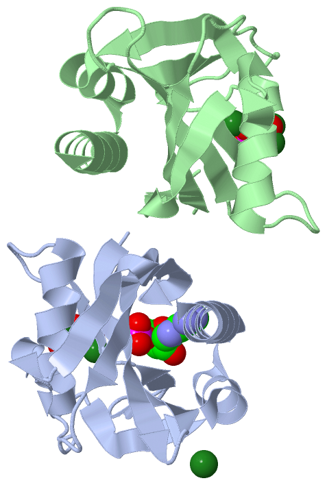
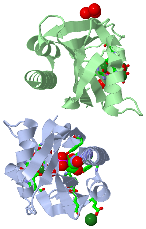
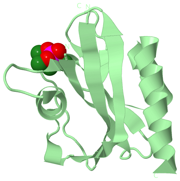
Asymmetric Unit (14, 14)
| No. | Name | Evidence | Residues | Description |
|---|
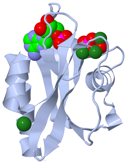
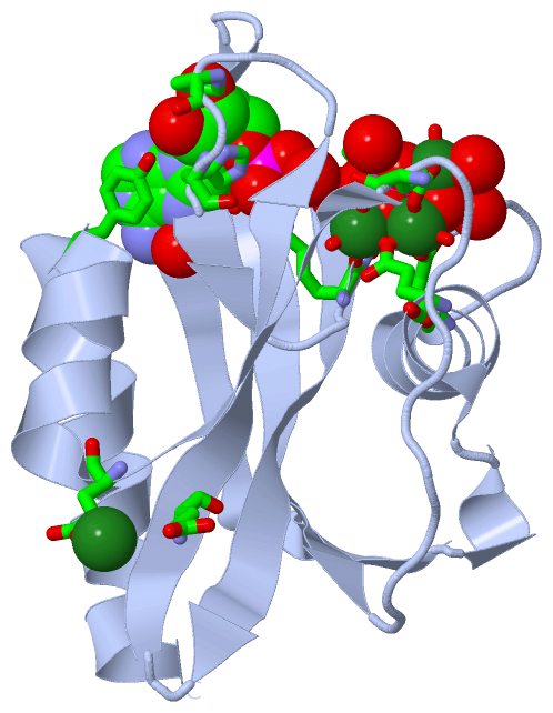
| 01 | AC1 | SOFTWARE | LYS A:36 , GLY A:37 , HIS A:38 , GLU A:52 , GLU A:56 , GLU A:103 , MG A:502 , MG A:503 , MG A:504 , MG A:505 , OH A:506 , HOH A:715 , HOH A:722 , HOH A:732 , HOH A:749 , HOH A:756 , HOH A:769 , HOH A:827 | BINDING SITE FOR RESIDUE PO4 A 402 |
| 02 | AC2 | SOFTWARE | LYS B:36 , GLY B:37 , HIS B:38 , GLU B:52 , GLU B:56 , GLU B:103 , MG B:601 , MG B:602 , MG B:603 , MG B:604 , OH B:605 , HOH B:615 , HOH B:631 , HOH B:635 , HOH B:654 , HOH B:669 , HOH B:672 , HOH B:703 | BINDING SITE FOR RESIDUE PO4 B 401 |
| 03 | AC3 | SOFTWARE | GLU A:52 , GLU A:103 , PO4 A:402 , MG A:503 , MG A:504 , MG A:505 , HOH A:708 , HOH A:742 , HOH A:752 , HOH A:756 | BINDING SITE FOR RESIDUE OH A 506 |
| 04 | AC4 | SOFTWARE | GLU B:52 , GLU B:103 , PO4 B:401 , MG B:601 , MG B:603 , MG B:604 , HOH B:611 , HOH B:633 , HOH B:650 , HOH B:654 | BINDING SITE FOR RESIDUE OH B 605 |
| 05 | AC5 | SOFTWARE | GLU A:19 , GLU A:111 , HOH A:729 , HOH A:737 , HOH B:616 , HOH B:638 , HOH B:644 | BINDING SITE FOR RESIDUE MG A 501 |
| 06 | AC6 | SOFTWARE | LYS A:36 , GLU A:56 , PO4 A:402 , MG A:505 , HOH A:722 , HOH A:732 , HOH A:734 | BINDING SITE FOR RESIDUE MG A 502 |
| 07 | AC7 | SOFTWARE | GLU A:52 , PO4 A:402 , MG A:504 , MG A:505 , OH A:506 , HOH A:715 , HOH A:752 , HOH A:756 | BINDING SITE FOR RESIDUE MG A 503 |
| 08 | AC8 | SOFTWARE | GLU A:103 , PO4 A:402 , MG A:503 , MG A:505 , OH A:506 , HOH A:742 , HOH A:749 , HOH A:756 | BINDING SITE FOR RESIDUE MG A 504 |
| 09 | AC9 | SOFTWARE | GLU A:52 , GLU A:56 , GLU A:103 , PO4 A:402 , MG A:502 , MG A:503 , MG A:504 , OH A:506 , HOH A:708 | BINDING SITE FOR RESIDUE MG A 505 |
| 10 | BC1 | SOFTWARE | GLU B:103 , PO4 B:401 , MG B:603 , MG B:604 , OH B:605 , HOH B:611 , HOH B:654 , HOH B:672 | BINDING SITE FOR RESIDUE MG B 601 |
| 11 | BC2 | SOFTWARE | LYS B:36 , GLU B:56 , PO4 B:401 , MG B:603 , HOH B:607 , HOH B:615 , HOH B:669 | BINDING SITE FOR RESIDUE MG B 602 |
| 12 | BC3 | SOFTWARE | GLU B:52 , GLU B:56 , GLU B:103 , PO4 B:401 , MG B:601 , MG B:602 , MG B:604 , OH B:605 , HOH B:650 | BINDING SITE FOR RESIDUE MG B 603 |
| 13 | BC4 | SOFTWARE | GLU B:52 , PO4 B:401 , MG B:601 , MG B:603 , OH B:605 , HOH B:633 , HOH B:635 , HOH B:654 | BINDING SITE FOR RESIDUE MG B 604 |
| 14 | BC5 | SOFTWARE | PRO A:29 , HIS A:31 , LYS A:36 , TYR A:76 , LYS A:83 , TYR A:121 , HOH A:782 , HOH A:843 | BINDING SITE FOR RESIDUE AMP A 701 |
|
 Description