Show PDB file:   
         Plain Text   HTML   (compressed file size)
QuickSearch:   
by PDB,NDB,UniProt,PROSITE Code or Search Term(s)  
(-)NMR Structure - model 1
(-)NMR Structure - all models
collapse expand < >
Image NMR Structure - model 1
NMR Structure - model 1  (Jmol Viewer)
Image NMR Structure - all models
NMR Structure - all models  (Jmol Viewer)

(-) Description

Title :  SOLUTION STRUCTURE OF THE VAM7P PX DOMAIN
 
Authors :  J. Lu, J. Garcia, I. Dulubova, T. C. Sudhof, J. Rizo
Date :  14 Dec 01  (Deposition) - 12 Jun 02  (Release) - 24 Feb 09  (Revision)
Method :  SOLUTION NMR
Resolution :  NOT APPLICABLE
Chains :  NMR Structure  :  A  (20x)
Keywords :  Px Domain, Vam7P, Phosphoinositide Binding, Endocytosis/Exocytosis Complex (Keyword Search: [Gene Ontology, PubMed, Web (Google))
 
Reference :  J. Lu, J. Garcia, I. Dulubova, T. C. Sudhof, J. Rizo
Solution Structure Of The Vam7P Px Domain.
Biochemistry V. 41 5956 2002
PubMed-ID: 11993989  |  Reference-DOI: 10.1021/BI020050B
(for further references see the PDB file header)

(-) Compounds

Molecule 1 - VACUOLAR MORPHOGENESIS PROTEIN VAM7
    ChainsA
    EngineeredYES
    Expression SystemESCHERICHIA COLI BL21
    Expression System PlasmidPGEX-KG
    Expression System StrainBL21
    Expression System Taxid511693
    Expression System Vector TypePLASMID
    FragmentPX DOMAIN, RESIDUES 8-124
    Organism CommonBAKER'S YEAST
    Organism ScientificSACCHAROMYCES CEREVISIAE
    Organism Taxid4932
    SynonymVAM7P

 Structural Features

(-) Chains, Units

  
NMR Structure (20x)

Summary Information (see also Sequences/Alignments below)

(-) Ligands, Modified Residues, Ions  (0, 0)

(no "Ligand,Modified Residues,Ions" information available for 1KMD)

(-) Sites  (0, 0)

(no "Site" information available for 1KMD)

(-) SS Bonds  (0, 0)

(no "SS Bond" information available for 1KMD)

(-) Cis Peptide Bonds  (0, 0)

(no "Cis Peptide Bond" information available for 1KMD)

 Sequence-Structure Mapping

(-) SAPs(SNPs)/Variants  (0, 0)

(no "SAP(SNP)/Variant" information available for 1KMD)

(-) PROSITE Motifs  (0, 0)

(no "PROSITE Motif" information available for 1KMD)

(-) Exons   (1, 1)

NMR Structure (1, 1)
 ENSEMBLUniProtKBPDB
No.Transcript IDExonExon IDGenome LocationLengthIDLocationLengthCountLocationLength
1.1YGL212W1YGL212W.1VII:91436-92386951VAM7_YEAST1-3163161A:8-124117

(-) Sequences/Alignments

NMR Structure
   Reformat: Number of residues per line =  ('0' or empty: single-line sequence representation)
  Number of residues per labelling interval =   
  UniProt sequence: complete  aligned part    
   Show mapping: SCOP domains CATH domains Pfam domains Secondary structure (by author)
SAPs(SNPs) PROSITE motifs Exons
(details for a mapped element are shown in a popup box when the mouse pointer rests over it)
Chain A from PDB  Type:PROTEIN  Length:117
 aligned with VAM7_YEAST | P32912 from UniProtKB/Swiss-Prot  Length:316

    Alignment length:117
                                    17        27        37        47        57        67        77        87        97       107       117       
           VAM7_YEAST     8 KMSEKLRIKVDDVKINPKYVLYGVSTPNKRLYKRYSEFWKLKTRLERDVGSTIPYDFPEKPGVLDRRWQRRYDDPEMIDERRIGLERFLNELYNDRFDSRWRDTKIAQDFLQLSKPN 124
               SCOP domains d1kmda_ A: Vam7p                                                                                                      SCOP domains
               CATH domains 1kmdA00 A:8-124 PX Domain                                                                                             CATH domains
               Pfam domains --PX-1kmdA01 A:10-121                                                                                             --- Pfam domains
         Sec.struct. author ........ee...ee....eeeeee.....eeehhhhhhhhhhhhhhhhh.......................hhhhhhhhhhhhhhhhhhhhh....hhhhhhhhhhhhh...... Sec.struct. author
                 SAPs(SNPs) --------------------------------------------------------------------------------------------------------------------- SAPs(SNPs)
                    PROSITE --------------------------------------------------------------------------------------------------------------------- PROSITE
               Transcript 1 Exon 1.1  PDB: A:8-124 UniProt: 1-316 [INCOMPLETE]                                                                    Transcript 1
                 1kmd A   8 KMSEKLRIKVDDVKINPKYVLYGVSTPNKRLYKRYSEFWKLKTRLERDVGSTIPYDFPEKPGVLDRRWQRRYDDPEMIDERRIGLERFLNELYNDRFDSRWRDTKIAQDFLQLSKPN 124
                                    17        27        37        47        57        67        77        87        97       107       117       

   Legend:   → Mismatch (orange background)
  - → Gap (green background, '-', border residues have a numbering label)
    → Modified Residue (blue background, lower-case, 'x' indicates undefined single-letter code, labelled with number + name)
  x → Chemical Group (purple background, 'x', labelled with number + name, e.g. ACE or NH2)
  extra numbering lines below/above indicate numbering irregularities and modified residue names etc., number ends below/above '|'

 Classification and Annotation

(-) SCOP Domains  (1, 1)

NMR Structure

(-) CATH Domains  (1, 1)

NMR Structure
(-)
Class: Alpha Beta (26913)

(-) Pfam Domains  (1, 1)

NMR Structure

(-) Gene Ontology  (16, 16)

NMR Structure(hide GO term definitions)
Chain A   (VAM7_YEAST | P32912)
molecular function
    GO:0005484    SNAP receptor activity    Acting as a marker to identify a membrane and interacting selectively with one or more SNAREs on another membrane to mediate membrane fusion.
    GO:0000149    SNARE binding    Interacting selectively and non-covalently with a SNARE (soluble N-ethylmaleimide-sensitive factor attached protein receptor) protein.
    GO:0035091    phosphatidylinositol binding    Interacting selectively and non-covalently with any inositol-containing glycerophospholipid, i.e. phosphatidylinositol (PtdIns) and its phosphorylated derivatives.
    GO:0032266    phosphatidylinositol-3-phosphate binding    Interacting selectively and non-covalently with phosphatidylinositol-3-phosphate, a derivative of phosphatidylinositol in which the inositol ring is phosphorylated at the 3' position.
    GO:0005515    protein binding    Interacting selectively and non-covalently with any protein or protein complex (a complex of two or more proteins that may include other nonprotein molecules).
biological process
    GO:0016236    macroautophagy    The major inducible pathway for the general turnover of cytoplasmic constituents in eukaryotic cells, it is also responsible for the degradation of active cytoplasmic enzymes and organelles during nutrient starvation. Macroautophagy involves the formation of double-membrane-bounded autophagosomes which enclose the cytoplasmic constituent targeted for degradation in a membrane-bounded structure. Autophagosomes then fuse with a lysosome (or vacuole) releasing single-membrane-bounded autophagic bodies that are then degraded within the lysosome (or vacuole). Though once thought to be a purely non-selective process, it appears that some types of macroautophagy, e.g. macropexophagy, macromitophagy, may involve selective targeting of the targets to be degraded.
    GO:0034727    piecemeal microautophagy of nucleus    Degradation of a cell nucleus by lysosomal microautophagy.
    GO:0032258    protein localization by the CVT pathway    A cytoplasm to vacuole targeting pathway that uses machinery common with autophagy. The CVT vesicle is formed when the receptor protein, Atg19, binds to the complexes of the target protein (aminopeptidase or alpha-mannosidase homododecamers), forming the Cvt complex. Atg11 binds to Atg9 and transports the CVT complex to the pre-autophagosome (PAS). The phagophore membrane expands around the CVT complex (excluding bulk cytoplasm) forming the CVT vesicle. This pathway is mostly observed in yeast.
    GO:0042144    vacuole fusion, non-autophagic    The fusion of two vacuole membranes to form a single vacuole.
    GO:0048278    vesicle docking    The initial attachment of a transport vesicle membrane to the target membrane, mediated by proteins protruding from the membrane of the vesicle and the target membrane. Docking requires only that the two membranes come close enough for these proteins to interact and adhere.
    GO:0006906    vesicle fusion    Fusion of the membrane of a transport vesicle with its target membrane.
cellular component
    GO:0031201    SNARE complex    A protein complex involved in membrane fusion; a stable ternary complex consisting of a four-helix bundle, usually formed from one R-SNARE and three Q-SNAREs with an ionic layer sandwiched between hydrophobic layers. One well-characterized example is the neuronal SNARE complex formed of synaptobrevin 2, syntaxin 1a, and SNAP-25.
    GO:0012505    endomembrane system    A collection of membranous structures involved in transport within the cell. The main components of the endomembrane system are endoplasmic reticulum, Golgi bodies, vesicles, cell membrane and nuclear envelope. Members of the endomembrane system pass materials through each other or though the use of vesicles.
    GO:0000329    fungal-type vacuole membrane    The lipid bilayer surrounding a vacuole, the shape of which correlates with cell cycle phase. The membrane separates its contents from the cytoplasm of the cell. An example of this structure is found in Saccharomyces cerevisiae.
    GO:0016021    integral component of membrane    The component of a membrane consisting of the gene products and protein complexes having at least some part of their peptide sequence embedded in the hydrophobic region of the membrane.
    GO:0005773    vacuole    A closed structure, found only in eukaryotic cells, that is completely surrounded by unit membrane and contains liquid material. Cells contain one or several vacuoles, that may have different functions from each other. Vacuoles have a diverse array of functions. They can act as a storage organelle for nutrients or waste products, as a degradative compartment, as a cost-effective way of increasing cell size, and as a homeostatic regulator controlling both turgor pressure and pH of the cytosol.
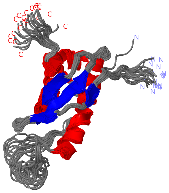
 Visualization

(-) Interactive Views

NMR Structure
  Complete Structure
    Jena3D(integrated viewing of ligand, site, SAP, PROSITE, SCOP information)
    WebMol | AstexViewer[tm]@PDBe
(Java Applets, require no local installation except for Java; loading may be slow)
    STRAP
(Java WebStart application, automatic local installation, requires Java; full application with system access!)
    RasMol
(require local installation)
    Molscript (VRML)
(requires installation of a VRML viewer; select preferred view via VRML and generate a mono or stereo PDF format file)
 
  Ligands, Modified Residues, Ions
(no "Ligands, Modified Residues, Ions" information available for 1kmd)
 
  Sites
(no "Sites" information available for 1kmd)
 
  Cis Peptide Bonds
(no "Cis Peptide Bonds" information available for 1kmd)
 

(-) Still Images

Jmol
  protein: cartoon or spacefill or dots and stick; nucleic acid: cartoon and stick; ligands: spacefill; active site: stick
Molscript
  protein, nucleic acid: cartoon; ligands: spacefill; active site: ball and stick

 Databases and Analysis Tools

(-) Databases

Access by PDB/NDB ID
  1kmd
    Family and Domain InformationProDom | SYSTERS
    General Structural InformationGlycoscienceDB | MMDB | NDB | OCA | PDB | PDBe | PDBj | PDBsum | PDBWiki | PQS | PROTEOPEDIA
    Orientation in MembranesOPM
    Protein SurfaceSURFACE
    Secondary StructureDSSP (structure derived) | HSSP (homology derived)
    Structural GenomicsGeneCensus
    Structural NeighboursCE | VAST
    Structure ClassificationCATH | Dali | SCOP
    Validation and Original DataBMRB Data View | BMRB Restraints Grid | EDS | PROCHECK | RECOORD | WHAT_CHECK
 
Access by UniProt ID/Accession number
  VAM7_YEAST | P32912
    Comparative Protein Structure ModelsModBase
    Genomic InformationEnsembl
    Protein-protein InteractionDIP
    Sequence, Family and Domain InformationInterPro | Pfam | SMART | UniProtKB/SwissProt
 
Access by Enzyme Classificator   (EC Number)
  (no 'Enzyme Classificator' available)
    General Enzyme InformationBRENDA | EC-PDB | Enzyme | IntEnz
    PathwayKEGG | MetaCyc
 
Access by Disease Identifier   (MIM ID)
  (no 'MIM ID' available)
    Disease InformationOMIM
 
Access by GenAge ID
  (no 'GenAge ID' available)
    Age Related InformationGenAge

(-) Analysis Tools

Access by PDB/NDB ID
    Domain InformationXDom
    Interatomic Contacts of Structural UnitsCSU
    Ligand-protein ContactsLPC
    Protein CavitiescastP
    Sequence and Secondary StructurePDBCartoon
    Structure AlignmentSTRAP(Java WebStart application, automatic local installation, requires Java; full application with system access!)
    Structure and Sequence BrowserSTING
 
Access by UniProt ID/Accession number
  VAM7_YEAST | P32912
    Protein Disorder PredictionDisEMBL | FoldIndex | GLOBPLOT (for more information see DisProt)

 Related Entries

(-) Entries Sharing at Least One Protein Chain (UniProt ID)

(no "Entries Sharing at Least One Protein Chain" available for 1KMD)

(-) Related Entries Specified in the PDB File

(no "Related Entries Specified in the PDB File" available for 1KMD)