| molecular function |
|---|
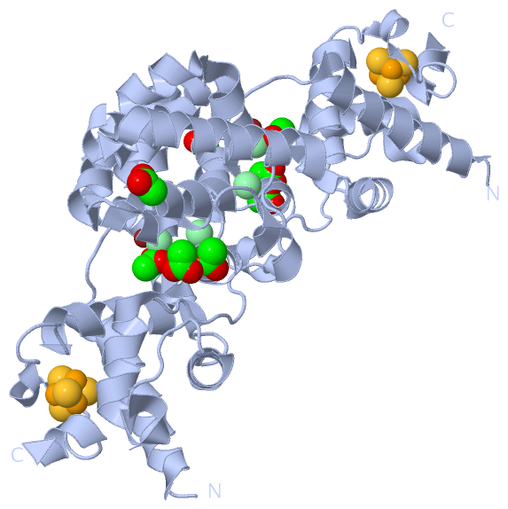
| | GO:0051539 | | 4 iron, 4 sulfur cluster binding | | Interacting selectively and non-covalently with a 4 iron, 4 sulfur (4Fe-4S) cluster; this cluster consists of four iron atoms, with the inorganic sulfur atoms found between the irons and acting as bridging ligands. |
| | GO:0003677 | | DNA binding | | Any molecular function by which a gene product interacts selectively and non-covalently with DNA (deoxyribonucleic acid). |
| | GO:0003906 | | DNA-(apurinic or apyrimidinic site) lyase activity | | Catalysis of the cleavage of the C-O-P bond 3' to the apurinic or apyrimidinic site in DNA by a beta-elimination reaction, leaving a 3'-terminal unsaturated sugar and a product with a terminal 5'-phosphate. |
| | GO:0003824 | | catalytic activity | | Catalysis of a biochemical reaction at physiological temperatures. In biologically catalyzed reactions, the reactants are known as substrates, and the catalysts are naturally occurring macromolecular substances known as enzymes. Enzymes possess specific binding sites for substrates, and are usually composed wholly or largely of protein, but RNA that has catalytic activity (ribozyme) is often also regarded as enzymatic. |
| | GO:0016787 | | hydrolase activity | | Catalysis of the hydrolysis of various bonds, e.g. C-O, C-N, C-C, phosphoric anhydride bonds, etc. Hydrolase is the systematic name for any enzyme of EC class 3. |
| | GO:0016798 | | hydrolase activity, acting on glycosyl bonds | | Catalysis of the hydrolysis of any glycosyl bond. |
| | GO:0051536 | | iron-sulfur cluster binding | | Interacting selectively and non-covalently with an iron-sulfur cluster, a combination of iron and sulfur atoms. |
| | GO:0046872 | | metal ion binding | | Interacting selectively and non-covalently with any metal ion. |
| biological process |
|---|
| | GO:0006281 | | DNA repair | | The process of restoring DNA after damage. Genomes are subject to damage by chemical and physical agents in the environment (e.g. UV and ionizing radiations, chemical mutagens, fungal and bacterial toxins, etc.) and by free radicals or alkylating agents endogenously generated in metabolism. DNA is also damaged because of errors during its replication. A variety of different DNA repair pathways have been reported that include direct reversal, base excision repair, nucleotide excision repair, photoreactivation, bypass, double-strand break repair pathway, and mismatch repair pathway. |
| | GO:0006284 | | base-excision repair | | In base excision repair, an altered base is removed by a DNA glycosylase enzyme, followed by excision of the resulting sugar phosphate. The small gap left in the DNA helix is filled in by the sequential action of DNA polymerase and DNA ligase. |
| | GO:0006974 | | cellular response to DNA damage stimulus | | Any process that results in a change in state or activity of a cell (in terms of movement, secretion, enzyme production, gene expression, etc.) as a result of a stimulus indicating damage to its DNA from environmental insults or errors during metabolism. |
| | GO:0008152 | | metabolic process | | The chemical reactions and pathways, including anabolism and catabolism, by which living organisms transform chemical substances. Metabolic processes typically transform small molecules, but also include macromolecular processes such as DNA repair and replication, and protein synthesis and degradation. |
 Description