Show PDB file:   
         Plain Text   HTML   (compressed file size)
QuickSearch:   
by PDB,NDB,UniProt,PROSITE Code or Search Term(s)  
(-)Asym./Biol. Unit
(-)Asym./Biol. Unit - sites
collapse expand < >
Image Asym./Biol. Unit
Asym./Biol. Unit  (Jmol Viewer)
Image Asym./Biol. Unit - sites
Asym./Biol. Unit - sites  (Jmol Viewer)

(-) Description

Title :  ENDOPOLYGALACTURONASE I FROM STEREUM PURPUREUM AT 0.96 A RESOLUTION
 
Authors :  T. Shimizu, T. Nakatsu, K. Miyairi, T. Okuno, H. Kato
Date :  10 Oct 01  (Deposition) - 05 Jun 02  (Release) - 13 Jul 11  (Revision)
Method :  X-RAY DIFFRACTION
Resolution :  0.96
Chains :  Asym./Biol. Unit :  A
Keywords :  Beta Helical Structure, Glycoside Hydrolase, Silver-Leaf Inducing Substance, Hydrolase (Keyword Search: [Gene Ontology, PubMed, Web (Google))
 
Reference :  T. Shimizu, T. Nakatsu, K. Miyairi, T. Okuno, H. Kato
Active-Site Architecture Of Endopolygalacturonase I From Stereum Purpureum Revealed By Crystal Structures In Native And Ligand-Bound Forms At Atomic Resolution.
Biochemistry V. 41 6651 2002
PubMed-ID: 12022868  |  Reference-DOI: 10.1021/BI025541A
(for further references see the PDB file header)

(-) Compounds

Molecule 1 - ENDOPOLYGALACTURONASE
    ChainsA
    EC Number3.2.1.15
    FragmentRESIDUES 1-335
    Organism ScientificCHONDROSTEREUM PURPUREUM
    Organism Taxid58369
    StrainASP-4B

 Structural Features

(-) Chains, Units

  1
Asymmetric/Biological Unit A

Summary Information (see also Sequences/Alignments below)
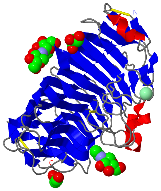
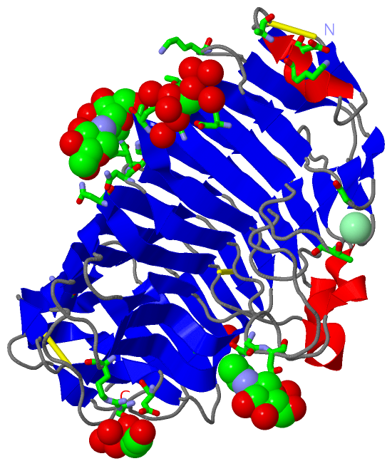
(-) Ligands, Modified Residues, Ions  (3, 5)

Asymmetric/Biological Unit (3, 5)
No.NameCountTypeFull Name
1CL1Ligand/IonCHLORIDE ION
2GOL2Ligand/IonGLYCEROL
3NAG2Ligand/IonN-ACETYL-D-GLUCOSAMINE

(-) Sites  (5, 5)

Asymmetric Unit (5, 5)
No.NameEvidenceResiduesDescription
1AC1SOFTWARELYS A:12 , ASP A:13 , ALA A:15 , ASN A:91 , ASN A:92 , HOH A:2213BINDING SITE FOR RESIDUE NAG A 2001
2AC2SOFTWAREALA A:129 , HIS A:130 , ASN A:160 , ASN A:161 , ASN A:182 , HOH A:2249 , HOH A:2470BINDING SITE FOR RESIDUE NAG A 2002
3AC3SOFTWAREGLY A:30 , THR A:55 , ASN A:308BINDING SITE FOR RESIDUE CL A 2003
4AC4SOFTWARELYS A:39 , ASP A:128 , ALA A:129 , ARG A:231 , ASP A:264 , VAL A:266 , LYS A:288 , HOH A:2082 , HOH A:2384BINDING SITE FOR RESIDUE GOL A 1001
5AC5SOFTWAREASP A:64 , THR A:66 , GLY A:105 , ASP A:128 , HOH A:2185 , HOH A:2215 , HOH A:2264 , HOH A:2293 , HOH A:2330 , HOH A:2390 , HOH A:2506BINDING SITE FOR RESIDUE GOL A 1002

(-) SS Bonds  (3, 3)

Asymmetric/Biological Unit
No.Residues
1A:3 -A:17
2A:175 -A:191
3A:300 -A:303

(-) Cis Peptide Bonds  (6, 6)

Asymmetric/Biological Unit
No.Residues
1Gly A:58 -Pro A:59
2His A:98 -Pro A:99
3Gly A:125 -Pro A:126
4Gly A:200 -Ser A:201
5Tyr A:262 -Pro A:263
6Gly A:331 -Gly A:332

 Sequence-Structure Mapping

(-) SAPs(SNPs)/Variants  (0, 0)

(no "SAP(SNP)/Variant" information available for 1K5C)

(-) PROSITE Motifs  (0, 0)

(no "PROSITE Motif" information available for 1K5C)

(-) Exons   (0, 0)

(no "Exon" information available for 1K5C)

(-) Sequences/Alignments

Asymmetric/Biological Unit
   Reformat: Number of residues per line =  ('0' or empty: single-line sequence representation)
  Number of residues per labelling interval =   
  UniProt sequence: complete  aligned part    
   Show mapping: SCOP domains CATH domains Pfam domains Secondary structure (by author)
SAPs(SNPs) PROSITE motifs Exons
(details for a mapped element are shown in a popup box when the mouse pointer rests over it)
Chain A from PDB  Type:PROTEIN  Length:333
 aligned with P79074_9AGAR | P79074 from UniProtKB/TrEMBL  Length:403

    Alignment length:333
                                    36        46        56        66        76        86        96       106       116       126       136       146       156       166       176       186       196       206       216       226       236       246       256       266       276       286       296       306       316       326       336       346       356   
         P79074_9AGAR    27 CTVKSVDDAKDIAGCSAVTLNGFTVPAGNTLVLNPDKGATVTMAGDITFAKTTLDGPLFTIDGTGINFVGADHIFDGNGALYWDGKGTNNGTHKPHPFLKIKGSGTYKKFEVLNSPAQAISVGPTDAHLTLDGITVDDFAGDTKNLGHNTDGFDVSANNVTIQNCIVKNQDDCIAINDGNNIRFENNQCSGGHGISIGSIATGKHVSNVVIKGNTVTRSMYGVRIKAQRTATSASVSGVTYDANTISGIAKYGVLISQSYPDDVGNPGTGAPFSDVNFTGGATTIKVNNAATRVTVECGNCSGNWNWSQLTVTGGKAGTIKSDKAKITGGQYL 359
               SCOP domains d1k5ca_ A: Polygalacturonase                                                                                                                                                                                                                                                                                                                  SCOP domains
               CATH domains 1k5cA00 A:3-335 Single-stranded right-handed beta-helix, Pectin lyase-like                                                                                                                                                                                                                                                                    CATH domains
               Pfam domains --------------Glyco_hydro_28-1k5cA01 A:17-331                                                                                                                                                                                                                                                                                            ---- Pfam domains
         Sec.struct. author .ee...hhhhhh....eeee..eee.....eee......eeee...eee.........eeeeeeeeeee....eee.hhhhh................eeeeeeeeee..eee......eeeeeeeeeeeee..eee.hhhhhhhh......eeee...eeee..eee.....eeeeeeeeeeee..eee....eeeeee....eeeeeeee..eeeeeeeeeeeeee......eeeeeeee..eeeeeeeeeeeeeeee............eeeeee....eeeee....eeeeee...ee.eeeeeeeeee......ee....eeeeee.. Sec.struct. author
                 SAPs(SNPs) --------------------------------------------------------------------------------------------------------------------------------------------------------------------------------------------------------------------------------------------------------------------------------------------------------------------------------------------- SAPs(SNPs)
                    PROSITE --------------------------------------------------------------------------------------------------------------------------------------------------------------------------------------------------------------------------------------------------------------------------------------------------------------------------------------------- PROSITE
                 Transcript --------------------------------------------------------------------------------------------------------------------------------------------------------------------------------------------------------------------------------------------------------------------------------------------------------------------------------------------- Transcript
                 1k5c A   3 CTVKSVDDAKDIAGCSAVTLNGFTVPAGNTLVLNPDKGATVTMAGDITFAKTTLDGPLFTIDGTGINFVGADHIFDGNGALYWDGKGTNNGTHKPHPFLKIKGSGTYKKFEVLNSPAQAISVGPTDAHLTLDGITVDDFAGDTKNLGHNTDGFDVSANNVTIQNCIVKNQDDCIAINDGNNIRFENNQCSGGHGISIGSIATGKHVSNVVIKGNTVTRSMYGVRIKAQRTATSASVSGVTYDANTISGIAKYGVLISQSYPDDVGNPGTGAPFSDVNFTGGATTIKVNNAATRVTVECGNCSGNWNWSQLTVTGGKAGTIKSDKAKITGGQYL 335
                                    12        22        32        42        52        62        72        82        92       102       112       122       132       142       152       162       172       182       192       202       212       222       232       242       252       262       272       282       292       302       312       322       332   

   Legend:   → Mismatch (orange background)
  - → Gap (green background, '-', border residues have a numbering label)
    → Modified Residue (blue background, lower-case, 'x' indicates undefined single-letter code, labelled with number + name)
  x → Chemical Group (purple background, 'x', labelled with number + name, e.g. ACE or NH2)
  extra numbering lines below/above indicate numbering irregularities and modified residue names etc., number ends below/above '|'

 Classification and Annotation

(-) SCOP Domains  (1, 1)

Asymmetric/Biological Unit

(-) CATH Domains  (1, 1)

Asymmetric/Biological Unit
(-)
Class: Mainly Beta (13760)

(-) Pfam Domains  (1, 1)

Asymmetric/Biological Unit

(-) Gene Ontology  (7, 7)

Asymmetric/Biological Unit(hide GO term definitions)
Chain A   (P79074_9AGAR | P79074)
molecular function
    GO:0016787    hydrolase activity    Catalysis of the hydrolysis of various bonds, e.g. C-O, C-N, C-C, phosphoric anhydride bonds, etc. Hydrolase is the systematic name for any enzyme of EC class 3.
    GO:0016798    hydrolase activity, acting on glycosyl bonds    Catalysis of the hydrolysis of any glycosyl bond.
    GO:0004650    polygalacturonase activity    Catalysis of the random hydrolysis of (1->4)-alpha-D-galactosiduronic linkages in pectate and other galacturonans.
biological process
    GO:0005975    carbohydrate metabolic process    The chemical reactions and pathways involving carbohydrates, any of a group of organic compounds based of the general formula Cx(H2O)y. Includes the formation of carbohydrate derivatives by the addition of a carbohydrate residue to another molecule.
    GO:0071555    cell wall organization    A process that results in the assembly, arrangement of constituent parts, or disassembly of the cell wall, the rigid or semi-rigid envelope lying outside the cell membrane of plant, fungal and most prokaryotic cells, maintaining their shape and protecting them from osmotic lysis.
    GO:0008152    metabolic process    The chemical reactions and pathways, including anabolism and catabolism, by which living organisms transform chemical substances. Metabolic processes typically transform small molecules, but also include macromolecular processes such as DNA repair and replication, and protein synthesis and degradation.
cellular component
    GO:0005576    extracellular region    The space external to the outermost structure of a cell. For cells without external protective or external encapsulating structures this refers to space outside of the plasma membrane. This term covers the host cell environment outside an intracellular parasite.

 Visualization

(-) Interactive Views

Asymmetric/Biological Unit
  Complete Structure
    Jena3D(integrated viewing of ligand, site, SAP, PROSITE, SCOP information)
    WebMol | AstexViewer[tm]@PDBe
(Java Applets, require no local installation except for Java; loading may be slow)
    STRAP
(Java WebStart application, automatic local installation, requires Java; full application with system access!)
    RasMol
(require local installation)
    Molscript (VRML)
(requires installation of a VRML viewer; select preferred view via VRML and generate a mono or stereo PDF format file)
 
  Ligands, Modified Residues, Ions
    CL  [ RasMol | Jena3D ]  +environment [ RasMol | Jena3D ]
    GOL  [ RasMol | Jena3D ]  +environment [ RasMol | Jena3D ]
    NAG  [ RasMol | Jena3D ]  +environment [ RasMol | Jena3D ]
 
  Sites
    AC1  [ RasMol ]  +environment [ RasMol ]
    AC2  [ RasMol ]  +environment [ RasMol ]
    AC3  [ RasMol ]  +environment [ RasMol ]
    AC4  [ RasMol ]  +environment [ RasMol ]
    AC5  [ RasMol ]  +environment [ RasMol ]
 
  Cis Peptide Bonds
    Gly A:125 - Pro A:126   [ RasMol ]  
    Gly A:200 - Ser A:201   [ RasMol ]  
    Gly A:331 - Gly A:332   [ RasMol ]  
    Gly A:58 - Pro A:59   [ RasMol ]  
    His A:98 - Pro A:99   [ RasMol ]  
    Tyr A:262 - Pro A:263   [ RasMol ]  
 

(-) Still Images

Jmol
  protein: cartoon or spacefill or dots and stick; nucleic acid: cartoon and stick; ligands: spacefill; active site: stick
Molscript
  protein, nucleic acid: cartoon; ligands: spacefill; active site: ball and stick

 Databases and Analysis Tools

(-) Databases

Access by PDB/NDB ID
  1k5c
    Family and Domain InformationProDom | SYSTERS
    General Structural InformationGlycoscienceDB | MMDB | NDB | OCA | PDB | PDBe | PDBj | PDBsum | PDBWiki | PQS | PROTEOPEDIA
    Orientation in MembranesOPM
    Protein SurfaceSURFACE
    Secondary StructureDSSP (structure derived) | HSSP (homology derived)
    Structural GenomicsGeneCensus
    Structural NeighboursCE | VAST
    Structure ClassificationCATH | Dali | SCOP
    Validation and Original DataBMRB Data View | BMRB Restraints Grid | EDS | PROCHECK | RECOORD | WHAT_CHECK
 
Access by UniProt ID/Accession number
  P79074_9AGAR | P79074
    Comparative Protein Structure ModelsModBase
    Genomic InformationEnsembl
    Protein-protein InteractionDIP
    Sequence, Family and Domain InformationInterPro | Pfam | SMART | UniProtKB/TrEMBL
 
Access by Enzyme Classificator   (EC Number)
  3.2.1.15
    General Enzyme InformationBRENDA | EC-PDB | Enzyme | IntEnz
    PathwayKEGG | MetaCyc
 
Access by Disease Identifier   (MIM ID)
  (no 'MIM ID' available)
    Disease InformationOMIM
 
Access by GenAge ID
  (no 'GenAge ID' available)
    Age Related InformationGenAge

(-) Analysis Tools

Access by PDB/NDB ID
    Domain InformationXDom
    Interatomic Contacts of Structural UnitsCSU
    Ligand-protein ContactsLPC
    Protein CavitiescastP
    Sequence and Secondary StructurePDBCartoon
    Structure AlignmentSTRAP(Java WebStart application, automatic local installation, requires Java; full application with system access!)
    Structure and Sequence BrowserSTING
 
Access by UniProt ID/Accession number
  P79074_9AGAR | P79074
    Protein Disorder PredictionDisEMBL | FoldIndex | GLOBPLOT (for more information see DisProt)

 Related Entries

(-) Entries Sharing at Least One Protein Chain (UniProt ID)

UniProtKB/TrEMBL
        P79074_9AGAR | P790741kcc 1kcd

(-) Related Entries Specified in the PDB File

1kcc 1KCC CONTAINS THE SAME ENZYME COMPLEXED WITH ONE GALACTURONATE.
1kcd 1KCD CONTAINS THE SAME ENZYME COMPLEXED WITH TWO GALACTURONATE.