Show PDB file:   
         Plain Text   HTML   (compressed file size)
QuickSearch:   
by PDB,NDB,UniProt,PROSITE Code or Search Term(s)  
(-)Asymmetric Unit
(-)Asym. Unit - sites
(-)Biological Unit 1
(-)Biol. Unit 1 - sites
collapse expand < >
Image Asymmetric Unit
Asymmetric Unit  (Jmol Viewer)
Image Asym. Unit - sites
Asym. Unit - sites  (Jmol Viewer)
Image Biological Unit 1
Biological Unit 1  (Jmol Viewer)
Image Biol. Unit 1 - sites
Biol. Unit 1 - sites  (Jmol Viewer)

(-) Description

Title :  CRYSTAL STRUCTURE OF 3,4-DIHYDROXY-2-BUTANONE 4-PHOSPHATE SYNTHASE (CATION FREE FORM)
 
Authors :  D. -I. Liao, Y. -J. Zheng, P. V. Viitanen, D. B. Jordan
Date :  06 Oct 01  (Deposition) - 06 Mar 02  (Release) - 16 Nov 11  (Revision)
Method :  X-RAY DIFFRACTION
Resolution :  1.50
Chains :  Asym. Unit :  A
Biol. Unit 1:  A  (2x)
Keywords :  Dihydroxybutanone Phosphate Synthase, Riboflavin Biosynthesis, Antimicrobial Target, Structure-Based Design, Isomerase (Keyword Search: [Gene Ontology, PubMed, Web (Google))
 
Reference :  D. I. Liao, Y. J. Zheng, P. V. Viitanen, D. B. Jordan
Structural Definition Of The Active Site And Catalytic Mechanism Of 3, 4-Dihydroxy-2-Butanone-4-Phosphate Synthase.
Biochemistry V. 41 1795 2002
PubMed-ID: 11827524  |  Reference-DOI: 10.1021/BI015652U

(-) Compounds

Molecule 1 - 3,4-DIHYDROXY-2-BUTANONE 4-PHOSPHATE SYNTHASE
    ChainsA
    EC Number5.4.99.-
    EngineeredYES
    Expression SystemESCHERICHIA COLI BL21(DE3)
    Expression System PlasmidPET-24A(+)
    Expression System StrainBL21(DE3)
    Expression System Taxid469008
    Expression System Vector TypePLASMID
    GeneRICE BLAST FUNGI
    Organism ScientificMAGNAPORTHE GRISEA
    Organism Taxid148305

 Structural Features

(-) Chains, Units

  1
Asymmetric Unit A
Biological Unit 1 (2x)A

Summary Information (see also Sequences/Alignments below)

(-) Ligands, Modified Residues, Ions  (1, 4)

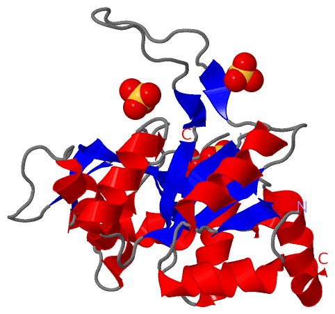
Asymmetric Unit (1, 4)
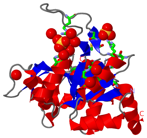
No.NameCountTypeFull Name
1SO44Ligand/IonSULFATE ION
Biological Unit 1 (1, 8)
No.NameCountTypeFull Name
1SO48Ligand/IonSULFATE ION

(-) Sites  (4, 4)

Asymmetric Unit (4, 4)
No.NameEvidenceResiduesDescription
1AC1SOFTWAREARG A:36 , THR A:92 , ARG A:150 , GLY A:152 , HIS A:153 , THR A:154 , HOH A:1112 , HOH A:1161BINDING SITE FOR RESIDUE SO4 A 1001
2AC2SOFTWAREGLN A:81 , ARG A:141 , ARG A:205 , HOH A:1080 , HOH A:1085 , HOH A:1175BINDING SITE FOR RESIDUE SO4 A 1002
3AC3SOFTWAREGLY A:192 , MET A:193 , ARG A:195 , HOH A:1070BINDING SITE FOR RESIDUE SO4 A 1003
4AC4SOFTWAREARG A:59 , ARG A:188 , HOH A:1014 , HOH A:1090 , HOH A:1135 , HOH A:1174 , HOH A:1224BINDING SITE FOR RESIDUE SO4 A 1004

(-) SS Bonds  (0, 0)

(no "SS Bond" information available for 1K49)

(-) Cis Peptide Bonds  (1, 1)

Asymmetric Unit
No.Residues
1Arg A:133 -Pro A:134

 Sequence-Structure Mapping

(-) SAPs(SNPs)/Variants  (0, 0)

(no "SAP(SNP)/Variant" information available for 1K49)

(-) PROSITE Motifs  (0, 0)

(no "PROSITE Motif" information available for 1K49)

(-) Exons   (0, 0)

(no "Exon" information available for 1K49)

(-) Sequences/Alignments

Asymmetric Unit
   Reformat: Number of residues per line =  ('0' or empty: single-line sequence representation)
  Number of residues per labelling interval =   
  UniProt sequence: complete  aligned part    
   Show mapping: SCOP domains CATH domains Pfam domains Secondary structure (by author)
SAPs(SNPs) PROSITE motifs Exons
(details for a mapped element are shown in a popup box when the mouse pointer rests over it)
Chain A from PDB  Type:PROTEIN  Length:208
 aligned with RIB3_MAGO7 | Q8TG90 from UniProtKB/Swiss-Prot  Length:233

    Alignment length:215
                                    20        30        40        50        60        70        80        90       100       110       120       130       140       150       160       170       180       190       200       210       220     
           RIB3_MAGO7    11 NFDAIPDVIQAFKNGEFVVVLDDPSRENEADLIIAAESVTTEQMAFMVRHSSGLICAPLTPERTTALDLPQMVTHNADPRGTAYTVSVDAEHPSTTTGISAHDRALACRMLAAPDAQPSHFRRPGHVFPLRAVAGGVRARRGHTEAGVELCRLAGKRPVAVISEIVDDGQEVEGRAVRAAPGMLRGDECVAFARRWGLKVCTIEDMIAHVEKTEG 225
               SCOP domains d1k49a_ A: 3,4-dihydroxy-2-butanone 4-phosphate synthase, DHBP synthase,        RibB                                                                                                                                    SCOP domains
               CATH domains 1k49A00 A:11-225 DHBP synthase                                                                                                                                                                                          CATH domains
               Pfam domains ----DHBP_synthase-1k49A01 A:15-220                                                                                                                                                                                ----- Pfam domains
         Sec.struct. author ...hhhhhhhhhhh...eeee........eeeeee....hhhhhhhhhhh.....eeeehhhhhhhhh.....-------.......eee.........hhhhhhhhhhhhhh...hhh.eeeeeeeeeee...hhhhh..hhhhhhhhhhhhh.....eeeee.ee..............ee.hhhhhhhhhhhh..eeeehhhhhhhhhhhhh Sec.struct. author
                 SAPs(SNPs) ----------------------------------------------------------------------------------------------------------------------------------------------------------------------------------------------------------------------- SAPs(SNPs)
                    PROSITE ----------------------------------------------------------------------------------------------------------------------------------------------------------------------------------------------------------------------- PROSITE
                 Transcript ----------------------------------------------------------------------------------------------------------------------------------------------------------------------------------------------------------------------- Transcript
                 1k49 A  11 NFDAIPDVIQAFKNGEFVVVLDDPSRENEADLIIAAESVTTEQMAFMVRHSSGLICAPLTPERTTALDLPQMV-------GTAYTVSVDAEHPSTTTGISAHDRALACRMLAAPDAQPSHFRRPGHVFPLRAVAGGVRARRGHTEAGVELCRLAGKRPVAVISEIVDDGQEVEGRAVRAAPGMLRGDECVAFARRWGLKVCTIEDMIAHVEKTEG 225
                                    20        30        40        50        60        70        80  |      -|      100       110       120       130       140       150       160       170       180       190       200       210       220     
                                                                                                   83      91                                                                                                                                      

   Legend:   → Mismatch (orange background)
  - → Gap (green background, '-', border residues have a numbering label)
    → Modified Residue (blue background, lower-case, 'x' indicates undefined single-letter code, labelled with number + name)
  x → Chemical Group (purple background, 'x', labelled with number + name, e.g. ACE or NH2)
  extra numbering lines below/above indicate numbering irregularities and modified residue names etc., number ends below/above '|'

 Classification and Annotation

(-) SCOP Domains  (1, 1)

Asymmetric Unit

(-) CATH Domains  (1, 1)

Asymmetric Unit
(-)
Class: Alpha Beta (26913)

(-) Pfam Domains  (1, 1)

Asymmetric Unit

(-) Gene Ontology  (5, 5)

Asymmetric Unit(hide GO term definitions)
Chain A   (RIB3_MAGO7 | Q8TG90)
molecular function
    GO:0008686    3,4-dihydroxy-2-butanone-4-phosphate synthase activity    Catalysis of the reaction: D-ribulose 5-phosphate = (2S)-2-hydroxy-3-oxobutyl phosphate + formate + H(+).
    GO:0016829    lyase activity    Catalysis of the cleavage of C-C, C-O, C-N and other bonds by other means than by hydrolysis or oxidation, or conversely adding a group to a double bond. They differ from other enzymes in that two substrates are involved in one reaction direction, but only one in the other direction. When acting on the single substrate, a molecule is eliminated and this generates either a new double bond or a new ring.
    GO:0046872    metal ion binding    Interacting selectively and non-covalently with any metal ion.
biological process
    GO:0009231    riboflavin biosynthetic process    The chemical reactions and pathways resulting in the formation of riboflavin (vitamin B2), the precursor for the coenzymes flavin mononucleotide (FMN) and flavin adenine dinucleotide (FAD).
cellular component
    GO:0005575    cellular_component    The part of a cell, extracellular environment or virus in which a gene product is located. A gene product may be located in one or more parts of a cell and its location may be as specific as a particular macromolecular complex, that is, a stable, persistent association of macromolecules that function together.

 Visualization

(-) Interactive Views

Asymmetric Unit
  Complete Structure
    Jena3D(integrated viewing of ligand, site, SAP, PROSITE, SCOP information)
    WebMol | AstexViewer[tm]@PDBe
(Java Applets, require no local installation except for Java; loading may be slow)
    STRAP
(Java WebStart application, automatic local installation, requires Java; full application with system access!)
    RasMol
(require local installation)
    Molscript (VRML)
(requires installation of a VRML viewer; select preferred view via VRML and generate a mono or stereo PDF format file)
 
  Ligands, Modified Residues, Ions
    SO4  [ RasMol | Jena3D ]  +environment [ RasMol | Jena3D ]
 
  Sites
    AC1  [ RasMol ]  +environment [ RasMol ]
    AC2  [ RasMol ]  +environment [ RasMol ]
    AC3  [ RasMol ]  +environment [ RasMol ]
    AC4  [ RasMol ]  +environment [ RasMol ]
 
  Cis Peptide Bonds
    Arg A:133 - Pro A:134   [ RasMol ]  
 
Biological Unit
  Complete Structure
    Biological Unit 1  [ Jena3D ]

(-) Still Images

Jmol
  protein: cartoon or spacefill or dots and stick; nucleic acid: cartoon and stick; ligands: spacefill; active site: stick
Molscript
  protein, nucleic acid: cartoon; ligands: spacefill; active site: ball and stick

 Databases and Analysis Tools

(-) Databases

Access by PDB/NDB ID
  1k49
    Family and Domain InformationProDom | SYSTERS
    General Structural InformationGlycoscienceDB | MMDB | NDB | OCA | PDB | PDBe | PDBj | PDBsum | PDBWiki | PQS | PROTEOPEDIA
    Orientation in MembranesOPM
    Protein SurfaceSURFACE
    Secondary StructureDSSP (structure derived) | HSSP (homology derived)
    Structural GenomicsGeneCensus
    Structural NeighboursCE | VAST
    Structure ClassificationCATH | Dali | SCOP
    Validation and Original DataBMRB Data View | BMRB Restraints Grid | EDS | PROCHECK | RECOORD | WHAT_CHECK
 
Access by UniProt ID/Accession number
  RIB3_MAGO7 | Q8TG90
    Comparative Protein Structure ModelsModBase
    Genomic InformationEnsembl
    Protein-protein InteractionDIP
    Sequence, Family and Domain InformationInterPro | Pfam | SMART | UniProtKB/SwissProt
 
Access by Enzyme Classificator   (EC Number)
  5.4.99.-
    General Enzyme InformationBRENDA | EC-PDB | Enzyme | IntEnz
    PathwayKEGG | MetaCyc
 
Access by Disease Identifier   (MIM ID)
  (no 'MIM ID' available)
    Disease InformationOMIM
 
Access by GenAge ID
  (no 'GenAge ID' available)
    Age Related InformationGenAge

(-) Analysis Tools

Access by PDB/NDB ID
    Domain InformationXDom
    Interatomic Contacts of Structural UnitsCSU
    Ligand-protein ContactsLPC
    Protein CavitiescastP
    Sequence and Secondary StructurePDBCartoon
    Structure AlignmentSTRAP(Java WebStart application, automatic local installation, requires Java; full application with system access!)
    Structure and Sequence BrowserSTING
 
Access by UniProt ID/Accession number
  RIB3_MAGO7 | Q8TG90
    Protein Disorder PredictionDisEMBL | FoldIndex | GLOBPLOT (for more information see DisProt)

 Related Entries

(-) Entries Sharing at Least One Protein Chain (UniProt ID)

UniProtKB/Swiss-Prot
        RIB3_MAGO7 | Q8TG901k4i 1k4l 1k4o 1k4p

(-) Related Entries Specified in the PDB File

(no "Related Entries Specified in the PDB File" available for 1K49)