|
|
|
|
 Description
|
|
Compounds
|
||||||||||||||||||||||||||||||||||||||||||||||||
Chains, Units
Summary Information (see also Sequences/Alignments below) |
Ligands, Modified Residues, Ions (1, 1)
NMR Structure (1, 1)
|
Sites (1, 1)
NMR Structure (1, 1)
|
SS Bonds (0, 0)| (no "SS Bond" information available for 1K3G) |
Cis Peptide Bonds (0, 0)| (no "Cis Peptide Bond" information available for 1K3G) |
SAPs(SNPs)/Variants (0, 0)| (no "SAP(SNP)/Variant" information available for 1K3G) |
PROSITE Motifs (0, 0)| (no "PROSITE Motif" information available for 1K3G) |
Exons (0, 0)| (no "Exon" information available for 1K3G) |
Sequences/Alignments
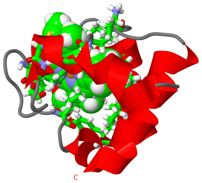
NMR StructureChain A from PDB Type:PROTEIN Length:71 aligned with CY553_SPOPA | P82599 from UniProtKB/Swiss-Prot Length:92 Alignment length:71 31 41 51 61 71 81 91 CY553_SPOPA 22 VDAEAVVQQKCISCHGGDLTGASAPAIDKAGANYSEEEILDIILNGQGGMPGGIAKGAEAEAVAAWLAEKK 92 SCOP domains d1k3ga_ A: Cytochrome c6 (synonym: cytochrome c553) SCOP domains CATH domains 1k3gA00 A:22-92 Cytochrome c CATH domains Pfam domains ----------------------------------------------------------------------- Pfam domains SAPs(SNPs) ----------------------------------------------------------------------- SAPs(SNPs) PROSITE ----------------------------------------------------------------------- PROSITE Transcript ----------------------------------------------------------------------- Transcript 1k3g A 22 VDAEAVVQQKCISCHGGDLTGASAPAIDKAGANYSEEEILDIILNGQGGMPGGIAKGAEAEAVAAWLAEKK 92 31 41 51 61 71 81 91
|
||||||||||||||||||||
SCOP Domains (1, 1)
NMR Structure
 |
CATH Domains (1, 1)
NMR Structure
|
Pfam Domains (0, 0)| (no "Pfam Domain" information available for 1K3G) |
Gene Ontology (8, 8)|
NMR Structure(hide GO term definitions) Chain A (CY553_SPOPA | P82599)
|
||||||||||||||||||||||||||||||||||||||||||||||||||||||||||||||||||
Interactive Views
|
||||||||||||||||||||||||||||||||||||||||||||||||||||||||||||||||||||||||||||||||||||||||||||||||||||||||||||||||||||||
Still Images
|
||||||||||||||||
Databases
|
||||||||||||||||||||||||||||||||||||||||||||||||||||||||||||||||||||||||||||||||||||||||||||||||||||||||||||||||||||||||||||||||||||||||||||||||||||||||||||||||
Analysis Tools
|
|||||||||||||||||||||||||||||||||||||||||||||||||||||||||||||
Entries Sharing at Least One Protein Chain (UniProt ID)
Related Entries Specified in the PDB File
|
|