Show PDB file:   
         Plain Text   HTML   (compressed file size)
QuickSearch:   
by PDB,NDB,UniProt,PROSITE Code or Search Term(s)  
(-)Asym./Biol. Unit - manually
(-)Asym./Biol. Unit
(-)Asym./Biol. Unit - sites
collapse expand < >
Image Asym./Biol. Unit - manually
Asym./Biol. Unit - manually  (Jmol Viewer)
Image Asym./Biol. Unit
Asym./Biol. Unit  (Jmol Viewer)
Image Asym./Biol. Unit - sites
Asym./Biol. Unit - sites  (Jmol Viewer)

(-) Description

Title :  MODIFIED FORM OF EOSINOPHIL-DERIVED NEUROTOXIN
 
Authors :  C. Chang, D. L. Newton, S. M. Rybak, A. Wlodawer
Date :  26 Sep 01  (Deposition) - 03 Apr 02  (Release) - 24 Feb 09  (Revision)
Method :  X-RAY DIFFRACTION
Resolution :  1.00
Chains :  Asym./Biol. Unit :  A
Keywords :  Rnase A Folding, Hydrolase (Keyword Search: [Gene Ontology, PubMed, Web (Google))
 
Reference :  C. Chang, D. L. Newton, S. M. Rybak, A. Wlodawer
Crystallographic And Functional Studies Of A Modified Form Of Eosinophil-Derived Neurotoxin (Edn) With Novel Biological Activities.
J. Mol. Biol. V. 317 119 2002
PubMed-ID: 11916383  |  Reference-DOI: 10.1006/JMBI.2002.5406
(for further references see the PDB file header)

(-) Compounds

Molecule 1 - EOSINOPHIL-DERIVED NEUROTOXIN
    ChainsA
    EC Number3.1.27.5
    EngineeredYES
    Expression SystemESCHERICHIA COLI
    Expression System Taxid562
    Organism CommonHUMAN
    Organism ScientificHOMO SAPIENS
    Organism Taxid9606
    SynonymRIBONUCLEASE 2

 Structural Features

(-) Chains, Units

  1
Asymmetric/Biological Unit A

Summary Information (see also Sequences/Alignments below)

(-) Ligands, Modified Residues, Ions  (1, 2)

Asymmetric/Biological Unit (1, 2)
No.NameCountTypeFull Name
1SO42Ligand/IonSULFATE ION

(-) Sites  (2, 2)

Asymmetric Unit (2, 2)
No.NameEvidenceResiduesDescription
1AC1SOFTWAREHIS A:-2 , GLN A:14 , HIS A:15 , HIS A:129 , LEU A:130 , HOH A:312 , HOH A:399 , HOH A:424 , HOH A:488 , HOH A:504BINDING SITE FOR RESIDUE SO4 A 301
2AC2SOFTWAREARG A:36 , LYS A:38 , ASN A:39 , GLN A:40 , HOH A:415 , HOH A:470 , HOH A:487BINDING SITE FOR RESIDUE SO4 A 302

(-) SS Bonds  (4, 4)

Asymmetric/Biological Unit
No.Residues
1A:23 -A:83
2A:37 -A:96
3A:55 -A:111
4A:62 -A:71

(-) Cis Peptide Bonds  (0, 0)

(no "Cis Peptide Bond" information available for 1K2A)

 Sequence-Structure Mapping

(-) SAPs(SNPs)/Variants  (2, 2)

Asymmetric/Biological Unit (2, 2)
  dbSNPPDB
No.SourceVariant IDVariantUniProt IDStatusIDChainVariant
1UniProtVAR_059820H100QRNAS2_HUMANPolymorphism8012891AH73Q
2UniProtVAR_013150H156NRNAS2_HUMANPolymorphism146887874AH129N

  SNP/SAP Summary Statistics (UniProtKB/Swiss-Prot)

(-) PROSITE Motifs  (1, 1)

Asymmetric/Biological Unit (1, 1)
 PROSITEUniProtKBPDB
No.IDACDescriptionIDLocationCountLocation
1RNASE_PANCREATICPS00127 Pancreatic ribonuclease family signature.RNAS2_HUMAN64-70  1A:37-43

(-) Exons   (1, 1)

Asymmetric/Biological Unit (1, 1)
 ENSEMBLUniProtKBPDB
No.Transcript IDExonExon IDGenome LocationLengthIDLocationLengthCountLocationLength
1.1ENST000003046251ENSE00001285128chr14:21423611-2142369585RNAS2_HUMAN-00--
1.2ENST000003046252ENSE00001124161chr14:21423926-21424595670RNAS2_HUMAN1-1631631A:-2-134136

(-) Sequences/Alignments

Asymmetric/Biological Unit
   Reformat: Number of residues per line =  ('0' or empty: single-line sequence representation)
  Number of residues per labelling interval =   
  UniProt sequence: complete  aligned part    
   Show mapping: SCOP domains CATH domains Pfam domains Secondary structure (by author)
SAPs(SNPs) PROSITE motifs Exons
(details for a mapped element are shown in a popup box when the mouse pointer rests over it)
Chain A from PDB  Type:PROTEIN  Length:136
 aligned with RNAS2_HUMAN | P10153 from UniProtKB/Swiss-Prot  Length:161

    Alignment length:136
                                    35        45        55        65        75        85        95       105       115       125       135       145       155      
          RNAS2_HUMAN    26 HVKPPQFTWAQWFETQHINMTSQQCTNAMQVINNYQRRCKNQNTFLLTTFANVVNVCGNPNMTCPSNKTRKNCHHSGSQVPLIHCNLTTPSPQNISNCRYAQTPANMFYIVACDNRDQRRDPPQYPVVPVHLDRII 161
               SCOP domains d1k2aa_ A: Eosinophil-derived neurotoxin (EDN)                                                                                           SCOP domains
               CATH domains 1k2aA00 A:-2-134 P-30 Protein                                                                                                            CATH domains
               Pfam domains -----RnaseA-1k2aA01 A:4-131                                                                                                          --- Pfam domains
         Sec.struct. author .......hhhhhhhhhhh.....hhhhhhhhhhhhhh....eeeee..hhhhhhhhh...............eee....eeeeeeeeee....hhhhheeeeeeeeeeeeeeeee...........eeeeeeeeee Sec.struct. author
                 SAPs(SNPs) --------------------------------------------------------------------------Q-------------------------------------------------------N----- SAPs(SNPs)
                    PROSITE --------------------------------------RNASE_P------------------------------------------------------------------------------------------- PROSITE
               Transcript 1 Exon 1.2  PDB: A:-2-134 UniProt: 1-163 [INCOMPLETE]                                                                                      Transcript 1
                 1k2a A  -2 HVKPPQFTWAQWFETQHINMTSQQCTNAMQVINNYQRRCKNQNTFLLTTFANVVNVCGNPNMTCPSNKTRKNCHHSGSQVPLIHCNLTTPSPQNISNCRYAQTPANMFYIVACDNRDQRRDPPQYPVVPVHLDRII 134
                             ||      8        18        28        38        48        58        68        78        88        98       108       118       128      
                             ||                                                                                                                                     
                            -1|                                                                                                                                     
                              1                                                                                                                                     

   Legend:   → Mismatch (orange background)
  - → Gap (green background, '-', border residues have a numbering label)
    → Modified Residue (blue background, lower-case, 'x' indicates undefined single-letter code, labelled with number + name)
  x → Chemical Group (purple background, 'x', labelled with number + name, e.g. ACE or NH2)
  extra numbering lines below/above indicate numbering irregularities and modified residue names etc., number ends below/above '|'

 Classification and Annotation

(-) SCOP Domains  (1, 1)

Asymmetric/Biological Unit

(-) CATH Domains  (1, 1)

Asymmetric/Biological Unit
(-)
Class: Alpha Beta (26913)

(-) Pfam Domains  (1, 1)

Asymmetric/Biological Unit

(-) Gene Ontology  (15, 15)

Asymmetric/Biological Unit(hide GO term definitions)
Chain A   (RNAS2_HUMAN | P10153)
molecular function
    GO:0004519    endonuclease activity    Catalysis of the hydrolysis of ester linkages within nucleic acids by creating internal breaks.
    GO:0016787    hydrolase activity    Catalysis of the hydrolysis of various bonds, e.g. C-O, C-N, C-C, phosphoric anhydride bonds, etc. Hydrolase is the systematic name for any enzyme of EC class 3.
    GO:0004518    nuclease activity    Catalysis of the hydrolysis of ester linkages within nucleic acids.
    GO:0003676    nucleic acid binding    Interacting selectively and non-covalently with any nucleic acid.
    GO:0004522    ribonuclease A activity    Catalysis of the endonucleolytic cleavage of RNA to 3'-phosphomononucleotides and 3'-phosphooligonucleotides ending in C-P or U-P with 2',3'-cyclic phosphate intermediates.
    GO:0004540    ribonuclease activity    Catalysis of the hydrolysis of phosphodiester bonds in chains of RNA.
biological process
    GO:0006401    RNA catabolic process    The chemical reactions and pathways resulting in the breakdown of RNA, ribonucleic acid, one of the two main type of nucleic acid, consisting of a long, unbranched macromolecule formed from ribonucleotides joined in 3',5'-phosphodiester linkage.
    GO:0090501    RNA phosphodiester bond hydrolysis    The RNA metabolic process in which the phosphodiester bonds between ribonucleotides are cleaved by hydrolysis.
    GO:0090502    RNA phosphodiester bond hydrolysis, endonucleolytic    The chemical reactions and pathways involving the hydrolysis of internal 3',5'-phosphodiester bonds in one or two strands of ribonucleotides.
    GO:0006935    chemotaxis    The directed movement of a motile cell or organism, or the directed growth of a cell guided by a specific chemical concentration gradient. Movement may be towards a higher concentration (positive chemotaxis) or towards a lower concentration (negative chemotaxis).
    GO:0090305    nucleic acid phosphodiester bond hydrolysis    The nucleic acid metabolic process in which the phosphodiester bonds between nucleotides are cleaved by hydrolysis.
    GO:1903955    positive regulation of protein targeting to mitochondrion    Any process that activates or increases the frequency, rate or extent of protein targeting to mitochondrion.
cellular component
    GO:0070062    extracellular exosome    A vesicle that is released into the extracellular region by fusion of the limiting endosomal membrane of a multivesicular body with the plasma membrane. Extracellular exosomes, also simply called exosomes, have a diameter of about 40-100 nm.
    GO:0005576    extracellular region    The space external to the outermost structure of a cell. For cells without external protective or external encapsulating structures this refers to space outside of the plasma membrane. This term covers the host cell environment outside an intracellular parasite.
    GO:0005764    lysosome    A small lytic vacuole that has cell cycle-independent morphology and is found in most animal cells and that contains a variety of hydrolases, most of which have their maximal activities in the pH range 5-6. The contained enzymes display latency if properly isolated. About 40 different lysosomal hydrolases are known and lysosomes have a great variety of morphologies and functions.
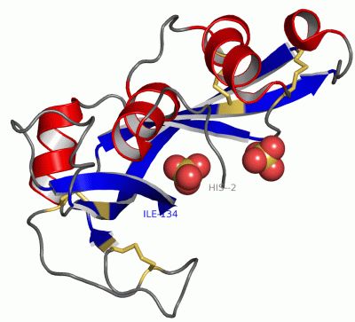
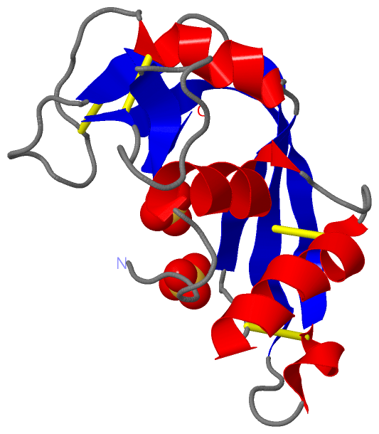
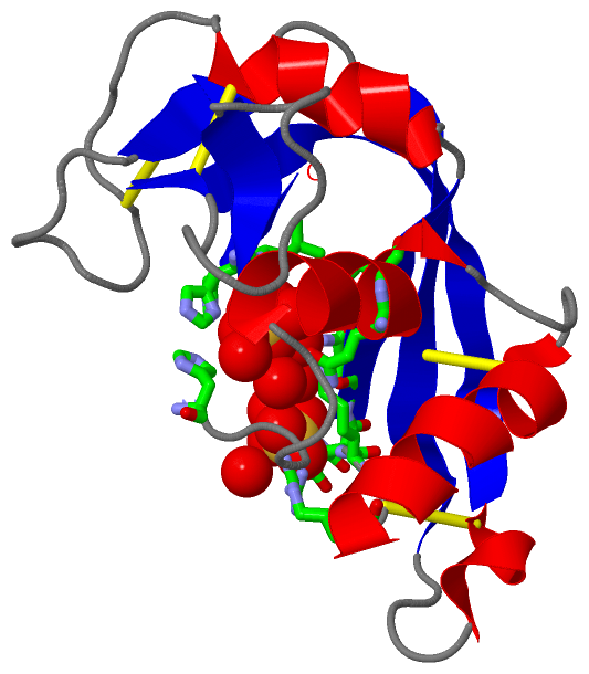
 Visualization

(-) Interactive Views

Asymmetric/Biological Unit
  Complete Structure
    Jena3D(integrated viewing of ligand, site, SAP, PROSITE, SCOP information)
    WebMol | AstexViewer[tm]@PDBe
(Java Applets, require no local installation except for Java; loading may be slow)
    STRAP
(Java WebStart application, automatic local installation, requires Java; full application with system access!)
    RasMol
(require local installation)
    Molscript (VRML)
(requires installation of a VRML viewer; select preferred view via VRML and generate a mono or stereo PDF format file)
 
  Ligands, Modified Residues, Ions
    SO4  [ RasMol | Jena3D ]  +environment [ RasMol | Jena3D ]
 
  Sites
    AC1  [ RasMol ]  +environment [ RasMol ]
    AC2  [ RasMol ]  +environment [ RasMol ]
 
  Cis Peptide Bonds
(no "Cis Peptide Bonds" information available for 1k2a)
 

(-) Still Images

Jmol
  protein: cartoon or spacefill or dots and stick; nucleic acid: cartoon and stick; ligands: spacefill; active site: stick
Molscript
  protein, nucleic acid: cartoon; ligands: spacefill; active site: ball and stick

 Databases and Analysis Tools

(-) Databases

Access by PDB/NDB ID
  1k2a
    Family and Domain InformationProDom | SYSTERS
    General Structural InformationGlycoscienceDB | MMDB | NDB | OCA | PDB | PDBe | PDBj | PDBsum | PDBWiki | PQS | PROTEOPEDIA
    Orientation in MembranesOPM
    Protein SurfaceSURFACE
    Secondary StructureDSSP (structure derived) | HSSP (homology derived)
    Structural GenomicsGeneCensus
    Structural NeighboursCE | VAST
    Structure ClassificationCATH | Dali | SCOP
    Validation and Original DataBMRB Data View | BMRB Restraints Grid | EDS | PROCHECK | RECOORD | WHAT_CHECK
 
Access by UniProt ID/Accession number
  RNAS2_HUMAN | P10153
    Comparative Protein Structure ModelsModBase
    Genomic InformationEnsembl
    Protein-protein InteractionDIP
    Sequence, Family and Domain InformationInterPro | Pfam | SMART | UniProtKB/SwissProt
 
Access by Enzyme Classificator   (EC Number)
  3.1.27.5
    General Enzyme InformationBRENDA | EC-PDB | Enzyme | IntEnz
    PathwayKEGG | MetaCyc
 
Access by Disease Identifier   (MIM ID)
  (no 'MIM ID' available)
    Disease InformationOMIM
 
Access by GenAge ID
  (no 'GenAge ID' available)
    Age Related InformationGenAge

(-) Analysis Tools

Access by PDB/NDB ID
    Domain InformationXDom
    Interatomic Contacts of Structural UnitsCSU
    Ligand-protein ContactsLPC
    Protein CavitiescastP
    Sequence and Secondary StructurePDBCartoon
    Structure AlignmentSTRAP(Java WebStart application, automatic local installation, requires Java; full application with system access!)
    Structure and Sequence BrowserSTING
 
Access by UniProt ID/Accession number
  RNAS2_HUMAN | P10153
    Protein Disorder PredictionDisEMBL | FoldIndex | GLOBPLOT (for more information see DisProt)

 Related Entries

(-) Entries Sharing at Least One Protein Chain (UniProt ID)

UniProtKB/Swiss-Prot
        RNAS2_HUMAN | P101531gqv 1hi2 1hi3 1hi4 1hi5 2bex 2bzz 2c01 2c02 2c05 5e13

(-) Related Entries Specified in the PDB File

(no "Related Entries Specified in the PDB File" available for 1K2A)