| molecular function |
|---|
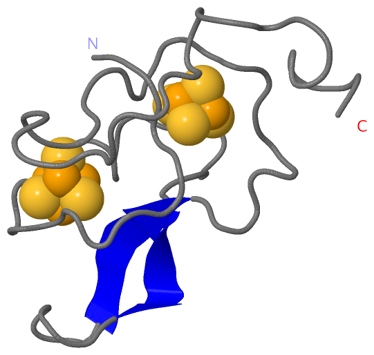
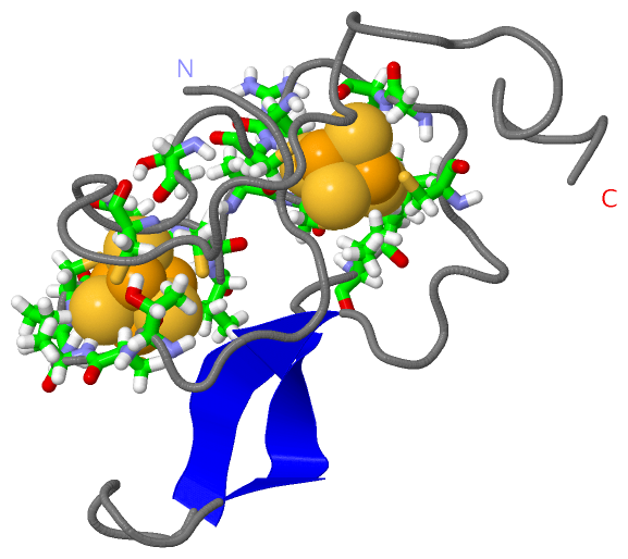
| | GO:0051539 | | 4 iron, 4 sulfur cluster binding | | Interacting selectively and non-covalently with a 4 iron, 4 sulfur (4Fe-4S) cluster; this cluster consists of four iron atoms, with the inorganic sulfur atoms found between the irons and acting as bridging ligands. |
| | GO:0009055 | | electron carrier activity | | Any molecular entity that serves as an electron acceptor and electron donor in an electron transport chain. An electron transport chain is a process in which a series of electron carriers operate together to transfer electrons from donors to any of several different terminal electron acceptors to generate a transmembrane electrochemical gradient. |
| | GO:0051536 | | iron-sulfur cluster binding | | Interacting selectively and non-covalently with an iron-sulfur cluster, a combination of iron and sulfur atoms. |
| | GO:0046872 | | metal ion binding | | Interacting selectively and non-covalently with any metal ion. |
| | GO:0016491 | | oxidoreductase activity | | Catalysis of an oxidation-reduction (redox) reaction, a reversible chemical reaction in which the oxidation state of an atom or atoms within a molecule is altered. One substrate acts as a hydrogen or electron donor and becomes oxidized, while the other acts as hydrogen or electron acceptor and becomes reduced. |
| biological process |
|---|
| | GO:0055114 | | oxidation-reduction process | | A metabolic process that results in the removal or addition of one or more electrons to or from a substance, with or without the concomitant removal or addition of a proton or protons. |
| | GO:0015979 | | photosynthesis | | The synthesis by organisms of organic chemical compounds, especially carbohydrates, from carbon dioxide (CO2) using energy obtained from light rather than from the oxidation of chemical compounds. |
| | GO:0009773 | | photosynthetic electron transport in photosystem I | | A photosynthetic electron transport chain in which electrons move from the primary electron acceptor (Quinone, X) through a chain of electron transport molecules in the thylakoid membrane until they reach ferredoxin which passes the electron to the ultimate electron acceptor; NADP. |
| cellular component |
|---|
| | GO:0016020 | | membrane | | A lipid bilayer along with all the proteins and protein complexes embedded in it an attached to it. |
| | GO:0009522 | | photosystem I | | A photosystem that contains an iron-sulfur reaction center associated with accessory pigments and electron carriers. In cyanobacteria and chloroplasts, photosystem I functions as a light-dependent plastocyanin-ferredoxin oxidoreductase, transferring electrons from plastocyanin to ferredoxin; in photosynthetic bacteria that have only a single type I photosystem, such as the green sulfur bacteria, electrons can go either to ferredoxin (Fd) -> NAD+ or to menaquinone (MK) -> Cytb/FeS -> Cytc555 -> photosystem I (cyclic photophosphorylation). |
| | GO:0009579 | | thylakoid | | A membranous cellular structure that bears the photosynthetic pigments in plants, algae, and cyanobacteria. In cyanobacteria thylakoids are of various shapes and are attached to, or continuous with, the plasma membrane. In eukaryotes they are flattened, membrane-bounded disk-like structures located in the chloroplasts; in the chloroplasts of higher plants the thylakoids form dense stacks called grana. Isolated thylakoid preparations can carry out photosynthetic electron transport and the associated phosphorylation. |
| | GO:0042651 | | thylakoid membrane | | The pigmented membrane of any thylakoid. |
 Description