Show PDB file:   
         Plain Text   HTML   (compressed file size)
QuickSearch:   
by PDB,NDB,UniProt,PROSITE Code or Search Term(s)  
(-)NMR Structure - manually
(-)NMR Structure - model 1
(-)NMR Structure - all models
collapse expand < >
Image NMR Structure - manually
NMR Structure - manually  (Jmol Viewer)
Image NMR Structure - model 1
NMR Structure - model 1  (Jmol Viewer)
Image NMR Structure - all models
NMR Structure - all models  (Jmol Viewer)

(-) Description

Title :  NMR STRUCTURE OF THE E. COLI PEPTIDYL-PROLYL CIS/TRANS-ISOMERASE PARVULIN 10
 
Authors :  A. Kuehlewein, G. Voll, B. Schelbert, H. Kessler, G. Fischer, J. U. Rahfeld, G. Gemmecker
Date :  25 Jul 01  (Deposition) - 17 Jun 03  (Release) - 24 Feb 09  (Revision)
Method :  SOLUTION NMR
Resolution :  NOT APPLICABLE
Chains :  NMR Structure  :  A  (18x)
Keywords :  Alpha-Beta Sandwich, Cis Peptide Bond, Isomerase (Keyword Search: [Gene Ontology, PubMed, Web (Google))
 
Reference :  A. Kuehlewein, G. Voll, B. H. Alvarez, H. Kessler, G. Fischer, J. U. Rahfeld, G. Gemmecker
Solution Structure Of Escherichia Coli Par10: The Prototypic Member Of The Parvulin Family Of Peptidyl-Prolyl Cis/Trans Isomerases.
Protein Sci. V. 13 2378 2004
PubMed-ID: 15322281  |  Reference-DOI: 10.1110/PS.04756704
(for further references see the PDB file header)

(-) Compounds

Molecule 1 - PEPTIDYL-PROLYL CIS-TRANS ISOMERASE C
    ChainsA
    EC Number5.2.1.8
    EngineeredYES
    Expression SystemESCHERICHIA COLI
    Expression System PlasmidPSEP612
    Expression System StrainM15[PREP4]
    Expression System Taxid562
    Expression System Vector TypePLASMID
    GenePARA
    Organism ScientificESCHERICHIA COLI
    Organism Taxid562
    SynonymPARVULIN, PPIASE C, ROTAMASE C

 Structural Features

(-) Chains, Units

  
NMR Structure (18x)

Summary Information (see also Sequences/Alignments below)

(-) Ligands, Modified Residues, Ions  (0, 0)

(no "Ligand,Modified Residues,Ions" information available for 1JNS)

(-) Sites  (0, 0)

(no "Site" information available for 1JNS)

(-) SS Bonds  (0, 0)

(no "SS Bond" information available for 1JNS)

(-) Cis Peptide Bonds  (1, 18)
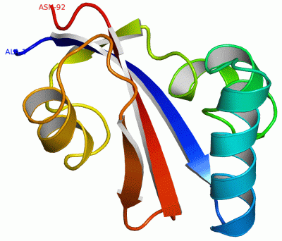
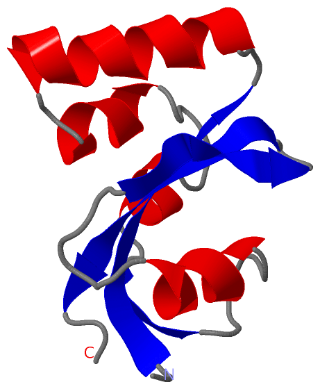
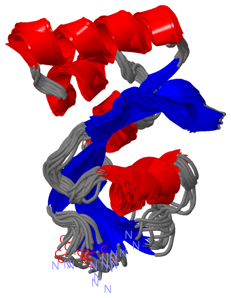
NMR Structure
No.ModelResidues
11, 2, 3, 4, 5, 6, 7, 8, 9, 10, 11, 12, 13, 14, 15, 16, 17, 18Gly A:75 -Pro A:76

 Sequence-Structure Mapping

(-) SAPs(SNPs)/Variants  (0, 0)

(no "SAP(SNP)/Variant" information available for 1JNS)

(-) PROSITE Motifs  (2, 2)

NMR Structure (2, 2)
 PROSITEUniProtKBPDB
No.IDACDescriptionIDLocationCountLocation
1PPIC_PPIASE_2PS50198 PpiC-type peptidyl-prolyl cis-trans isomerase family profile.PPIC_ECOLI2-91  1A:1-90
2PPIC_PPIASE_1PS01096 PpiC-type peptidyl-prolyl cis-trans isomerase signature.PPIC_ECOLI31-51  1A:30-50

(-) Exons   (0, 0)

(no "Exon" information available for 1JNS)

(-) Sequences/Alignments

NMR Structure
   Reformat: Number of residues per line =  ('0' or empty: single-line sequence representation)
  Number of residues per labelling interval =   
  UniProt sequence: complete  aligned part    
   Show mapping: SCOP domains CATH domains Pfam domains Secondary structure (by author)
SAPs(SNPs) PROSITE motifs Exons
(details for a mapped element are shown in a popup box when the mouse pointer rests over it)
Chain A from PDB  Type:PROTEIN  Length:92
 aligned with PPIC_ECOLI | P0A9L5 from UniProtKB/Swiss-Prot  Length:93

    Alignment length:92
                                    11        21        31        41        51        61        71        81        91  
            PPIC_ECOLI    2 AKTAAALHILVKEEKLALDLLEQIKNGADFGKLAKKHSICPSGKRGGDLGEFRQGQMVPAFDKVVFSCPVLEPTGPLHTQFGYHIIKVLYRN 93
               SCOP domains d1jnsa_ A: Parvulin 10 (rotamase C)                                                          SCOP domains
               CATH domains 1jnsA00 A:1-92  [code=3.10.50.40, no name defined]                                           CATH domains
               Pfam domains -------Rotamase-1jnsA01 A:8-90                                                            -- Pfam domains
         Sec.struct. author ..eeeeeeeee..hhhhhhhhhhhhhh..hhhhhhh......hhhhh..eeee......hhhhhhhh......eeeeee..eeeeeeee... Sec.struct. author
                 SAPs(SNPs) -------------------------------------------------------------------------------------------- SAPs(SNPs)
                PROSITE (1) PPIC_PPIASE_2  PDB: A:1-90 UniProt: 2-91                                                  -- PROSITE (1)
                PROSITE (2) -----------------------------PPIC_PPIASE_1        ------------------------------------------ PROSITE (2)
                 Transcript -------------------------------------------------------------------------------------------- Transcript
                  1jns A  1 AKTAAALHILVKEEKLALDLLEQIKNGADFGKLAKKHSICPSGKRGGDLGEFRQGQMVPAFDKVVFSCPVLEPTGPLHTQFGYHIIKVLYRN 92
                                    10        20        30        40        50        60        70        80        90  

   Legend:   → Mismatch (orange background)
  - → Gap (green background, '-', border residues have a numbering label)
    → Modified Residue (blue background, lower-case, 'x' indicates undefined single-letter code, labelled with number + name)
  x → Chemical Group (purple background, 'x', labelled with number + name, e.g. ACE or NH2)
  extra numbering lines below/above indicate numbering irregularities and modified residue names etc., number ends below/above '|'

 Classification and Annotation

(-) SCOP Domains  (1, 1)

NMR Structure

(-) CATH Domains  (1, 1)

NMR Structure
(-)
Class: Alpha Beta (26913)

(-) Pfam Domains  (1, 1)

NMR Structure
(-)
Clan: FKBP (43)

(-) Gene Ontology  (5, 5)

NMR Structure(hide GO term definitions)
Chain A   (PPIC_ECOLI | P0A9L5)
molecular function
    GO:0016853    isomerase activity    Catalysis of the geometric or structural changes within one molecule. Isomerase is the systematic name for any enzyme of EC class 5.
    GO:0003755    peptidyl-prolyl cis-trans isomerase activity    Catalysis of the reaction: peptidyl-proline (omega=180) = peptidyl-proline (omega=0).
    GO:0005515    protein binding    Interacting selectively and non-covalently with any protein or protein complex (a complex of two or more proteins that may include other nonprotein molecules).
biological process
    GO:0000413    protein peptidyl-prolyl isomerization    The modification of a protein by cis-trans isomerization of a proline residue.
cellular component
    GO:0005737    cytoplasm    All of the contents of a cell excluding the plasma membrane and nucleus, but including other subcellular structures.

 Visualization

(-) Interactive Views

NMR Structure
  Complete Structure
    Jena3D(integrated viewing of ligand, site, SAP, PROSITE, SCOP information)
    WebMol | AstexViewer[tm]@PDBe
(Java Applets, require no local installation except for Java; loading may be slow)
    STRAP
(Java WebStart application, automatic local installation, requires Java; full application with system access!)
    RasMol
(require local installation)
    Molscript (VRML)
(requires installation of a VRML viewer; select preferred view via VRML and generate a mono or stereo PDF format file)
 
  Ligands, Modified Residues, Ions
(no "Ligands, Modified Residues, Ions" information available for 1jns)
 
  Sites
(no "Sites" information available for 1jns)
 
  Cis Peptide Bonds
    Gly A:75 - Pro A:76   [ RasMol ]  
 

(-) Still Images

Jmol
  protein: cartoon or spacefill or dots and stick; nucleic acid: cartoon and stick; ligands: spacefill; active site: stick
Molscript
  protein, nucleic acid: cartoon; ligands: spacefill; active site: ball and stick

 Databases and Analysis Tools

(-) Databases

Access by PDB/NDB ID
  1jns
    Family and Domain InformationProDom | SYSTERS
    General Structural InformationGlycoscienceDB | MMDB | NDB | OCA | PDB | PDBe | PDBj | PDBsum | PDBWiki | PQS | PROTEOPEDIA
    Orientation in MembranesOPM
    Protein SurfaceSURFACE
    Secondary StructureDSSP (structure derived) | HSSP (homology derived)
    Structural GenomicsGeneCensus
    Structural NeighboursCE | VAST
    Structure ClassificationCATH | Dali | SCOP
    Validation and Original DataBMRB Data View | BMRB Restraints Grid | EDS | PROCHECK | RECOORD | WHAT_CHECK
 
Access by UniProt ID/Accession number
  PPIC_ECOLI | P0A9L5
    Comparative Protein Structure ModelsModBase
    Genomic InformationEnsembl
    Protein-protein InteractionDIP
    Sequence, Family and Domain InformationInterPro | Pfam | SMART | UniProtKB/SwissProt
 
Access by Enzyme Classificator   (EC Number)
  5.2.1.8
    General Enzyme InformationBRENDA | EC-PDB | Enzyme | IntEnz
    PathwayKEGG | MetaCyc
 
Access by Disease Identifier   (MIM ID)
  (no 'MIM ID' available)
    Disease InformationOMIM
 
Access by GenAge ID
  (no 'GenAge ID' available)
    Age Related InformationGenAge

(-) Analysis Tools

Access by PDB/NDB ID
    Domain InformationXDom
    Interatomic Contacts of Structural UnitsCSU
    Ligand-protein ContactsLPC
    Protein CavitiescastP
    Sequence and Secondary StructurePDBCartoon
    Structure AlignmentSTRAP(Java WebStart application, automatic local installation, requires Java; full application with system access!)
    Structure and Sequence BrowserSTING
 
Access by UniProt ID/Accession number
  PPIC_ECOLI | P0A9L5
    Protein Disorder PredictionDisEMBL | FoldIndex | GLOBPLOT (for more information see DisProt)

 Related Entries

(-) Entries Sharing at Least One Protein Chain (UniProt ID)

UniProtKB/Swiss-Prot
        PPIC_ECOLI | P0A9L51jnt

(-) Related Entries Specified in the PDB File

1jnt SAME PROTEIN, MINIMIZED AVERAGE STRUCTURE