|
|
|
|
 Description
|
|
Compounds
|
||||||||||||||||||||||||||||||||||||||||||||||||
Chains, Units
Summary Information (see also Sequences/Alignments below) |
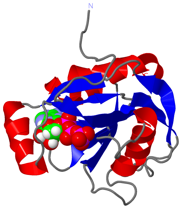
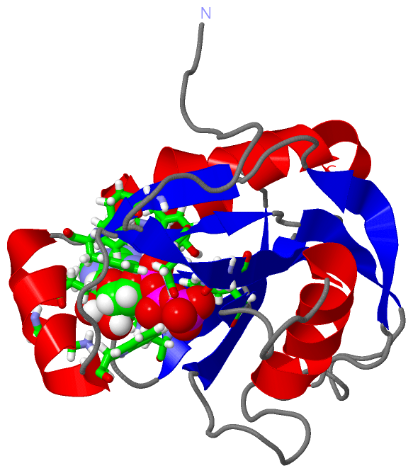
Ligands, Modified Residues, Ions (1, 1)
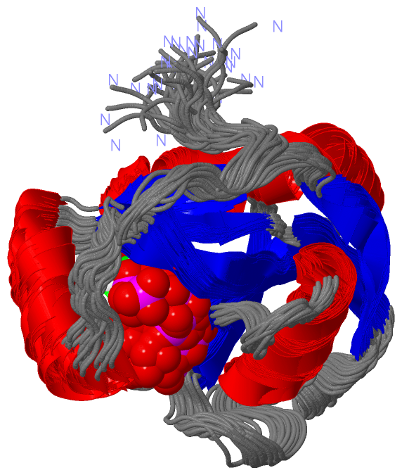
NMR Structure (1, 1)
|
Sites (1, 1)
NMR Structure (1, 1)
|
SS Bonds (0, 0)| (no "SS Bond" information available for 1JKN) |
Cis Peptide Bonds (0, 0)| (no "Cis Peptide Bond" information available for 1JKN) |
SAPs(SNPs)/Variants (0, 0)| (no "SAP(SNP)/Variant" information available for 1JKN) |
PROSITE Motifs (0, 0)| (no "PROSITE Motif" information available for 1JKN) |
Exons (0, 0)| (no "Exon" information available for 1JKN) |
Sequences/Alignments
NMR StructureChain A from PDB Type:PROTEIN Length:165 aligned with O04841_LUPAN | O04841 from UniProtKB/TrEMBL Length:199 Alignment length:191 18 28 38 48 58 68 78 88 98 108 118 128 138 148 158 168 178 188 198 O04841_LUPAN 9 SPPTNFHFRKYPSKFLKFSSLSLAFRYCHSSMDSPPEGYRRNVGICLMNNDKKIFAASRLDIPDAWQMPQGGIDEGEDPRNAAIRELREETGVTSAEVIAEVPYWLTYDFPPKVREKLNIQWGSDWKGQAQKWFLFKFTGQDQEINLLGDGSEKPEFGEWSWVTPEQLIDLTVEFKKPVYKEVLSVFAPHL 199 SCOP domains d1 jkna_ A: Diadenosine tetraphosphate hydrolase (Ap4A hydrolase) SCOP domains CATH domains 1j knA00 A:1-165 Nucleoside Triphosphate Pyrophosphohydrolase CATH domains Pfam domains --------------------------------------NUDIX-1jknA01 A:13-160 ----- Pfam domains SAPs(SNPs) ----------------------------------------------------------------------------------------------------------------------------------------------------------------------------------------------- SAPs(SNPs) PROSITE ----------------------------------------------------------------------------------------------------------------------------------------------------------------------------------------------- PROSITE Transcript ----------------------------------------------------------------------------------------------------------------------------------------------------------------------------------------------- Transcript 1jkn A 1 GP--------------------------LGSMDSPPEGYRRNVGICLMNNDKKIFAASRLDIPDAWQMPQGGIDEGEDPRNAAIRELREETGVTSAEVIAEVPYWLTYDFPPKVREKLNIQWGSDWKGQAQKWFLFKFTGQDQEINLLGDGSEKPEFGEWSWVTPEQLIDLTVEFKKPVYKEVLSVFAPHL 165 | - - |4 14 24 34 44 54 64 74 84 94 104 114 124 134 144 154 164 2 3
|
||||||||||||||||||||
SCOP Domains (1, 1)
NMR Structure
 |
CATH Domains (1, 1)
NMR Structure
|
Pfam Domains (1, 1)
NMR Structure
|
Gene Ontology (4, 4)|
NMR Structure(hide GO term definitions) Chain A (O04841_LUPAN | O04841)
|
||||||||||||||||||||||||||||||
Interactive Views
|
||||||||||||||||||||||||||||||||||||||||||||||||||||||||||||||||||||||||||||||||||||||||||||||||||||||||||||||||||||||
Still Images
|
||||||||||||||||
Databases
|
||||||||||||||||||||||||||||||||||||||||||||||||||||||||||||||||||||||||||||||||||||||||||||||||||||||||||||||||||||||||||||||||||||||||||||||||||||||||||||||||
Analysis Tools
|
|||||||||||||||||||||||||||||||||||||||||||||||||||||||||||||
Entries Sharing at Least One Protein Chain (UniProt ID)
Related Entries Specified in the PDB File
|
|