|
|
|
|
 Description
|
|
Compounds
|
||||||||||||||||||||||||||||||||
Chains, Units
Summary Information (see also Sequences/Alignments below) |
Ligands, Modified Residues, Ions (2, 5)| Asymmetric/Biological Unit (2, 5) |
Sites (5, 5)
Asymmetric Unit (5, 5)
|
SS Bonds (0, 0)| (no "SS Bond" information available for 1J84) |
Cis Peptide Bonds (0, 0)| (no "Cis Peptide Bond" information available for 1J84) |
SAPs(SNPs)/Variants (0, 0)| (no "SAP(SNP)/Variant" information available for 1J84) |
PROSITE Motifs (0, 0)| (no "PROSITE Motif" information available for 1J84) |
Exons (0, 0)| (no "Exon" information available for 1J84) |
Sequences/Alignments
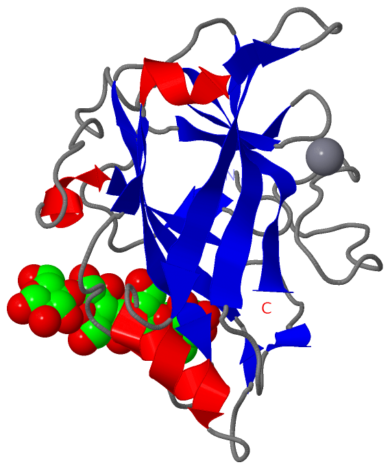
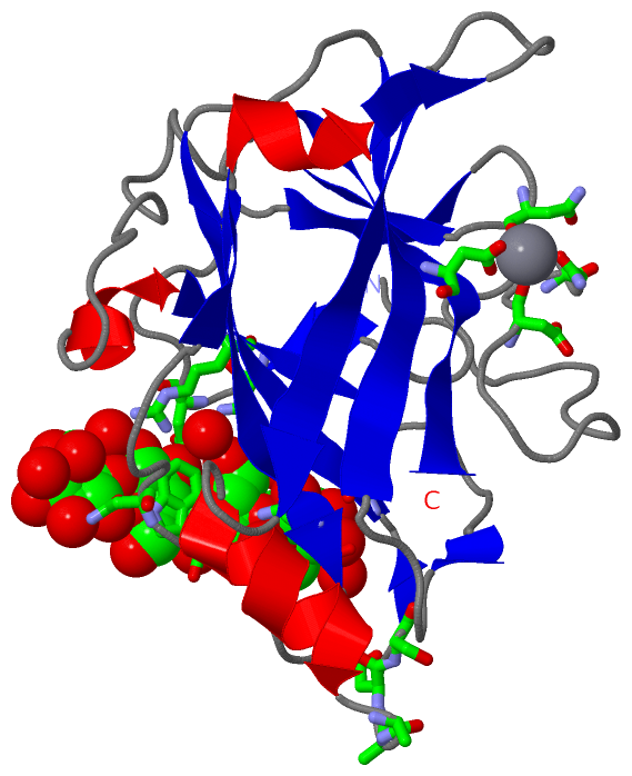
Asymmetric/Biological UnitChain A from PDB Type:PROTEIN Length:179 aligned with P94622_CLOCL | P94622 from UniProtKB/TrEMBL Length:557 Alignment length:179 387 397 407 417 427 437 447 457 467 477 487 497 507 517 527 537 547 P94622_CLOCL 378 QPTAPKDFSSGFWDFNDGTTQGFGVNPDSPITAINVENANNALKISNLNSKGSNDLSEGNFWANVRISADIWGQSINIYGDTKLTMDVIAPTPVNVSIAAIPQSSTHGWGNPTRAIRVWTNNFVAQTDGTYKATLTISTNDSPNFNTIATDAADSVVTNMILFVGSNSDNISLDNIKFT 556 SCOP domains d1j84a_ A: Endo-1,4-beta glucanase EngF SCOP domains CATH domains 1j84A00 A:27-205 Galactose-binding domain-like CATH domains Pfam domains ----------------------------------------------------------------------------------------------------------------------------------------------------------------------------------- Pfam domains SAPs(SNPs) ----------------------------------------------------------------------------------------------------------------------------------------------------------------------------------- SAPs(SNPs) PROSITE ----------------------------------------------------------------------------------------------------------------------------------------------------------------------------------- PROSITE Transcript ----------------------------------------------------------------------------------------------------------------------------------------------------------------------------------- Transcript 1j84 A 27 QPTAPKDFSSGFWDFNDGTTQGFGVNPDSPITAINVENANNALKISNLNSKGSNDLSEGNFWANVRISADIWGQSINIYGDTKLTMDVIAPTPVNVSIAAIPQSSTHGWGNPTRAIRVWTNNFVAQTDGTYKATLTISTNDSPNFNTIATDAADSVVTNMILFVGSNSDNISLDNIKFT 205 36 46 56 66 76 86 96 106 116 126 136 146 156 166 176 186 196
|
||||||||||||||||||||
SCOP Domains (1, 1)
Asymmetric/Biological Unit
 |
CATH Domains (1, 1)
Asymmetric/Biological Unit
|
Pfam Domains (0, 0)| (no "Pfam Domain" information available for 1J84) |
Gene Ontology (8, 8)|
Asymmetric/Biological Unit(hide GO term definitions) Chain A (P94622_CLOCL | P94622)
|
||||||||||||||||||||||||||||||||||||||||||||||||||||||||||||
Interactive Views
|
|||||||||||||||||||||||||||||||||||||||||||||||||||||||||||||||||||||||||||||||||||||||||||||||||||||||||||||||||||||||||||||||||||||||||||||||||||||||||
Still Images
|
||||||||||||||||
Databases
|
||||||||||||||||||||||||||||||||||||||||||||||||||||||||||||||||||||||||||||||||||||||||||||||||||||||||||||||||||||||||||||||||||||||||||||||||||||||||||||||||
Analysis Tools
|
|||||||||||||||||||||||||||||||||||||||||||||||||||||||||||||
Entries Sharing at Least One Protein Chain (UniProt ID)
Related Entries Specified in the PDB File
|
|