|
|
|
|
 Description
|
|
Compounds
|
||||||||||||||||||||||||||||||||||||||||||||||||||||||||||||||||||||||||||||||||
Chains, Units
Summary Information (see also Sequences/Alignments below) |
Ligands, Modified Residues, Ions (2, 22)| Asymmetric/Biological Unit (2, 22) |
Sites (10, 10)
Asymmetric Unit (10, 10)
|
SS Bonds (8, 8)
Asymmetric/Biological Unit
|
||||||||||||||||||||||||||||||||||||
Cis Peptide Bonds (0, 0)| (no "Cis Peptide Bond" information available for 1J35) |
SAPs(SNPs)/Variants (0, 0)| (no "SAP(SNP)/Variant" information available for 1J35) |
PROSITE Motifs (4, 6)
Asymmetric/Biological Unit (4, 6)
|
||||||||||||||||||||||||||||||||||||||||||||||||||||||||||||||||
Exons (2, 2)
Asymmetric/Biological Unit (2, 2)
|
||||||||||||||||||||||||||||||||||||||||||||||||||||||||||||||||||||||||||||||||||||||||||||||||||||||||||||||||||||||||
Sequences/Alignments
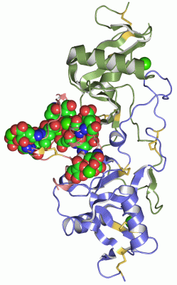
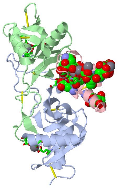
Asymmetric/Biological UnitChain A from PDB Type:PROTEIN Length:129 aligned with SL9A_PROFL | P23806 from UniProtKB/Swiss-Prot Length:152 Alignment length:129 33 43 53 63 73 83 93 103 113 123 133 143 SL9A_PROFL 24 DCLSGWSSYEGHCYKAFEKYKTWEDAERVCTEQAKGAHLVSIESSGEADFVAQLVTQNMKRLDFYIWIGLRVQGKVKQCNSEWSDGSSVSYENWIEAESKTCLGLEKETDFRKWVNIYCGQQNPFVCEA 152 SCOP domains d1j35a_ A: Snake coagglutinin alpha chain SCOP domains CATH domains 1j35A00 A:1-129 Mannose-Binding Protein A, subunit A CATH domains Pfam domains --------------------------------------------------------------------------------------------------------------------------------- Pfam domains SAPs(SNPs) --------------------------------------------------------------------------------------------------------------------------------- SAPs(SNPs) PROSITE (2) --------------------------------------------------------------------------------------------------------------------------------- PROSITE (2) PROSITE (3) --------C_TYPE_LECTIN_2 PDB: A:9-128 UniProt: 32-151 - PROSITE (3) PROSITE (4) --------------------------------------------------------------------------------------------------------------------------------- PROSITE (4) PROSITE (5) --------------------------------------------------------------------------------------------------------------------------------- PROSITE (5) PROSITE (6) -----------------------------------------------------------------------------------------------------C_TYPE_LECTIN_1 -- PROSITE (6) Transcript --------------------------------------------------------------------------------------------------------------------------------- Transcript 1j35 A 1 DCPSGWSSYEGHCYKPFKLYKTWDDAERFCTEQAKGGHLVSIESAGEADFVAQLVTENIQNTKSYVWIGLRVQGKEKQCSSEWSDGSSVSYENWIEAESKTCLGLEKETGFRKWVNIYCGQQNPFVCEA 129 10 20 30 40 50 60 70 80 90 100 110 120 Chain A from PDB Type:PROTEIN Length:129 aligned with SLA_PROFL | Q7LZ71 from UniProtKB/Swiss-Prot Length:129 Alignment length:129 10 20 30 40 50 60 70 80 90 100 110 120 SLA_PROFL 1 DCPSGWSSYEGHCYKPFKLYKTWDDAERFCTEQAKGGHLVSIESAGEADFVAQLVTENIQNTKSYVWIGLRVQGKEKQCSSEWSDGSSVSYENWIEAESKTCLGLEKETGFRKWVNIYCGQQNPFVCEA 129 SCOP domains d1j35a_ A: Snake coagglutinin alpha chain SCOP domains CATH domains 1j35A00 A:1-129 Mannose-Binding Protein A, subunit A CATH domains Pfam domains --------------------------------------------------------------------------------------------------------------------------------- Pfam domains SAPs(SNPs) --------------------------------------------------------------------------------------------------------------------------------- SAPs(SNPs) PROSITE (1) --------C_TYPE_LECTIN_2 PDB: A:9-128 UniProt: 9-128 - PROSITE (1) PROSITE (2) --------------------------------------------------------------------------------------------------------------------------------- PROSITE (2) PROSITE (3) --------------------------------------------------------------------------------------------------------------------------------- PROSITE (3) PROSITE (4) -----------------------------------------------------------------------------------------------------C_TYPE_LECTIN_1 -- PROSITE (4) Transcript --------------------------------------------------------------------------------------------------------------------------------- Transcript 1j35 A 1 DCPSGWSSYEGHCYKPFKLYKTWDDAERFCTEQAKGGHLVSIESAGEADFVAQLVTENIQNTKSYVWIGLRVQGKEKQCSSEWSDGSSVSYENWIEAESKTCLGLEKETGFRKWVNIYCGQQNPFVCEA 129 10 20 30 40 50 60 70 80 90 100 110 120 Chain B from PDB Type:PROTEIN Length:123 aligned with SL9B_PROFL | P23807 from UniProtKB/Swiss-Prot Length:146 Alignment length:123 33 43 53 63 73 83 93 103 113 123 133 143 SL9B_PROFL 24 DCPSDWSSYEGHCYKPFSEPKNWADAENFCTQQHAGGHLVSFQSSEEADFVVKLAFQTFGHSIFWMGLSNVWNQCNWQWSNAAMLRYKAWAEESYCVYFKSTNNKWRSRACRMMAQFVCEFQA 146 SCOP domains d1j35b_ B: Snake coagglutinin beta chain SCOP domains CATH domains 1j35B00 B:201-323 Mannose-Binding Protein A, subunit A CATH domains Pfam domains --------------------------------------------------------------------------------------------------------------------------- Pfam domains SAPs(SNPs) --------------------------------------------------------------------------------------------------------------------------- SAPs(SNPs) PROSITE (2) --------C_TYPE_LECTIN_2 PDB: B:209-320 UniProt: 32-143 --- PROSITE (2) PROSITE (3) --------------------------------------------------------------------------------------------------------------------------- PROSITE (3) PROSITE (4) --------------------------------------------------------------------------------------------------------------------------- PROSITE (4) PROSITE (5) -----------------------------------------------------------------------------------------------C_TYPE_LECTIN_1 ---- PROSITE (5) Transcript --------------------------------------------------------------------------------------------------------------------------- Transcript 1j35 B 201 DCPSDWSSYEGHCYKPFSEPKNWADAENFCTQQHAGGHLVSFQSSEEADFVVKLAFQTFGHSIFWMGLSNVWNQCNWQWSNAAMLRYKAWAEESYCVYFKSTNNKWRSRACRMMAQFVCEFQA 323 210 220 230 240 250 260 270 280 290 300 310 320 Chain C from PDB Type:PROTEIN Length:46 aligned with FA9_BOVIN | P00741 from UniProtKB/Swiss-Prot Length:462 Alignment length:46 56 66 76 86 FA9_BOVIN 47 YNSGKLEEFVRGNLERECKEEKCSFEEAREVFENTEKTTEFWKQYV 92 SCOP domains d1j35c_ C: Coagulation factor IX (IXa) SCOP domains CATH domains 1j35C00 C:401-446 Coagulation Factor IX CATH domains Pfam domains ---------------------------------------------- Pfam domains SAPs(SNPs) ---------------------------------------------- SAPs(SNPs) PROSITE (2) ---------------------------------------------- PROSITE (2) PROSITE (3) ---------------------------------------------- PROSITE (3) PROSITE (4) GLA_2 PDB: C:401-446 UniProt: 47-93 PROSITE (4) PROSITE (5) ----------------GLA_1 PDB: C:417-442 ---- PROSITE (5) Transcript 1 Exon 1.2 PDB: C:401-438 [INCOMPLETE] Exon 1.3 Transcript 1 1j35 C 401 YNSGKLeeFVRGNLeReCKeeKCSFeeAReVFeNTeKTTeFWKQYV 446 |410 | |420| ||430 | | 440 || | | || || | | | | 407-CGU | | || || | | | | 408-CGU | | || || | | | | 415-CGU|| || | | | | 417-CGU || | | | | 420-CGU || | | | | 421-CGU|| | | | | 426-CGU | | | 427-CGU | | |
|
||||||||||||||||||||
SCOP Domains (3, 3)
Asymmetric/Biological Unit
 |
CATH Domains (2, 3)
Asymmetric/Biological Unit
|
Pfam Domains (0, 0)| (no "Pfam Domain" information available for 1J35) |
Gene Ontology (15, 23)|
Asymmetric/Biological Unit(hide GO term definitions) Chain A (SL9A_PROFL | P23806)
Chain A (SLA_PROFL | Q7LZ71)
Chain B (SL9B_PROFL | P23807)
Chain C (FA9_BOVIN | P00741)
|
||||||||||||||||||||||||||||||||||||||||||||||||||||||||||||||||||||||||||||||||||||||||||||||||||||||||||||||||||||||||||||||||||||||||||||||||||||||||||||||||||||||||||||||||||||||||||||||||
Interactive Views
|
||||||||||||||||||||||||||||||||||||||||||||||||||||||||||||||||||||||||||||||||||||||||||||||||||||||||||||||||||||||||||||||||||||||||||||||||||||||||||||||||||||||||||||||||||||||||||||
Still Images
|
||||||||||||||||
Databases
|
||||||||||||||||||||||||||||||||||||||||||||||||||||||||||||||||||||||||||||||||||||||||||||||||||||||||||||||||||||||||||||||||||||||||||||||||||||||||||||||||||||||||||||||||||||||||||||||||||||||||||||||||||||||||||||||||||||||||||||||
Analysis Tools
|
||||||||||||||||||||||||||||||||||||||||||||||||||||||||||||||||||||||||||||||||||||||||||||||
Entries Sharing at Least One Protein Chain (UniProt ID)
Related Entries Specified in the PDB File
|
|