|
|
|
|
 Description
|
|
Compounds
|
||||||||||||||||||||||||
Chains, Units
Summary Information (see also Sequences/Alignments below) |
Ligands, Modified Residues, Ions (2, 2)| Asymmetric/Biological Unit (2, 2) |
Sites (2, 2)
Asymmetric Unit (2, 2)
|
SS Bonds (1, 1)
Asymmetric/Biological Unit
|
||||||||
Cis Peptide Bonds (1, 1)
Asymmetric/Biological Unit
|
||||||||
SAPs(SNPs)/Variants (0, 0)| (no "SAP(SNP)/Variant" information available for 1J1T) |
PROSITE Motifs (0, 0)| (no "PROSITE Motif" information available for 1J1T) |
Exons (0, 0)| (no "Exon" information available for 1J1T) |
Sequences/Alignments
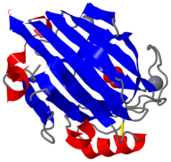
Asymmetric/Biological UnitChain A from PDB Type:PROTEIN Length:228 aligned with P84143_ALTS2 | P84143 from UniProtKB/TrEMBL Length:233 Alignment length:228 15 25 35 45 55 65 75 85 95 105 115 125 135 145 155 165 175 185 195 205 215 225 P84143_ALTS2 6 STIPSSITSGSIFDLEGDNPNPLVDDSTLVFVPLEAQHITPNGNGWRHEYKVKESLRVAMTQTYEVFEATVKVEMSDGGKTIISQHHASDTGTISKVYVSDTDESGFNDSVANNGIFDVYVRLRNTSGNEEKFALGTMTSGETFNLRVVNNYGDVEVTAFGNSFGIPVEDDSQSYFKFGNYLQSQDPYTLDKCGEAGNSNSFKNCFEDLGITESKVTMTNVTYTRETN 233 SCOP domains d1j1ta_ A: Alginate lyase SCOP domains CATH domains 1j1tA00 A:6-233 [code=2.60.120.200, no name defined] CATH domains Pfam domains ------------------------------------------------------------------------------------------------------------------------------------------------------------------------------------------------------------------------------------ Pfam domains SAPs(SNPs) ------------------------------------------------------------------------------------------------------------------------------------------------------------------------------------------------------------------------------------ SAPs(SNPs) PROSITE ------------------------------------------------------------------------------------------------------------------------------------------------------------------------------------------------------------------------------------ PROSITE Transcript ------------------------------------------------------------------------------------------------------------------------------------------------------------------------------------------------------------------------------------ Transcript 1j1t A 6 STIPSSITSGSIFDLEGDNPNPLVDDSTLVFVPLEAQHITPNGNGWRHEYKVKESLRVAMTQTYEVFEATVKVEMSDGGKTIISQHHASDTGTISKVYVSDTDESGFNDSVANNGIFDVYVRLRNTSGNEEKFALGTMTSGETFNLRVVNNYGDVEVTAFGNSFGIPVEDDSQSYFKFGNYLQSQDPYTLDKCGEAGNSNSFKNCFEDLGITESKVTMTNVTYTRETN 233 15 25 35 45 55 65 75 85 95 105 115 125 135 145 155 165 175 185 195 205 215 225
|
||||||||||||||||||||
SCOP Domains (1, 1)
Asymmetric/Biological Unit
 |
CATH Domains (1, 1)
Asymmetric/Biological Unit
|
Pfam Domains (0, 0)| (no "Pfam Domain" information available for 1J1T) |
Gene Ontology (1, 1)|
Asymmetric/Biological Unit(hide GO term definitions) Chain A (P84143_ALTS2 | P84143)
|
||||||||||||
Interactive Views
|
|||||||||||||||||||||||||||||||||||||||||||||||||||||||||||||||||||||||||||||||||||||||||||||||||||||||||||||||||||||||||||||||||||||
Still Images
|
||||||||||||||||
Databases
|
||||||||||||||||||||||||||||||||||||||||||||||||||||||||||||||||||||||||||||||||||||||||||||||||||||||||||||||||||||||||||||||||||||||||||||||||||||||||||||||||
Analysis Tools
|
|||||||||||||||||||||||||||||||||||||||||||||||||||||||||||||
Entries Sharing at Least One Protein Chain (UniProt ID)
Related Entries Specified in the PDB File
|
|