Show PDB file:   
         Plain Text   HTML   (compressed file size)
QuickSearch:   
by PDB,NDB,UniProt,PROSITE Code or Search Term(s)  
(-)NMR Structure - model 1
(-)NMR Structure - model 1, sites
(-)NMR Structure - all models
collapse expand < >
Image NMR Structure - model 1
NMR Structure - model 1  (Jmol Viewer)
Image NMR Structure - model 1, sites
NMR Structure - model 1, sites  (Jmol Viewer)
Image NMR Structure - all models
NMR Structure - all models  (Jmol Viewer)

(-) Description

Title :  GLUCOSE PERMEASE (DOMAIN IIB), NMR, 11 STRUCTURES
 
Authors :  M. Eberstadt, S. G. Grdadolnik, G. Gemmecker, H. Kessler, A. Buhr, B. Erni
Date :  23 Mar 96  (Deposition) - 14 Oct 96  (Release) - 24 Feb 09  (Revision)
Method :  SOLUTION NMR
Resolution :  NOT APPLICABLE
Chains :  NMR Structure  :  A  (11x)
Keywords :  Phosphotransferase System, Sugar Transport, Transferase, Phosphorylation, Transmembrane, Inner Membrane, Phoshphotransferase (Keyword Search: [Gene Ontology, PubMed, Web (Google))
 
Reference :  M. Eberstadt, S. G. Grdadolnik, G. Gemmecker, H. Kessler, A. Buhr, B. Erni
Solution Structure Of The Iib Domain Of The Glucose Transporter Of Escherichia Coli.
Biochemistry V. 35 11286 1996
PubMed-ID: 8784182  |  Reference-DOI: 10.1021/BI960492L
(for further references see the PDB file header)

(-) Compounds

Molecule 1 - GLUCOSE PERMEASE
    ChainsA
    EC Number2.7.1.69
    EngineeredYES
    Expression SystemESCHERICHIA COLI
    Expression System GenePTSG
    Expression System PlasmidPQBH
    Expression System Taxid562
    FragmentDOMAIN IIB
    Organism ScientificESCHERICHIA COLI
    Organism Taxid562
    StrainK-12, W3110

 Structural Features

(-) Chains, Units

  
NMR Structure (11x)

Summary Information (see also Sequences/Alignments below)

(-) Ligands, Modified Residues, Ions  (0, 0)

(no "Ligand,Modified Residues,Ions" information available for 1IBA)

(-) Sites  (1, 1)

NMR Structure (1, 1)
No.NameEvidenceResiduesDescription
1S1UNKNOWNCYS A:35PHOSPHORYLATION SITE CYS 35.

(-) SS Bonds  (0, 0)

(no "SS Bond" information available for 1IBA)

(-) Cis Peptide Bonds  (0, 0)

(no "Cis Peptide Bond" information available for 1IBA)

 Sequence-Structure Mapping

(-) SAPs(SNPs)/Variants  (0, 0)

(no "SAP(SNP)/Variant" information available for 1IBA)

(-) PROSITE Motifs  (1, 1)

NMR Structure (1, 1)
 PROSITEUniProtKBPDB
No.IDACDescriptionIDLocationCountLocation
1PTS_EIIB_TYPE_1_CYSPS01035 PTS EIIB domains cysteine phosphorylation site signature.PTGCB_ECOLI414-431  1A:28-45

(-) Exons   (0, 0)

(no "Exon" information available for 1IBA)

(-) Sequences/Alignments

NMR Structure
   Reformat: Number of residues per line =  ('0' or empty: single-line sequence representation)
  Number of residues per labelling interval =   
  UniProt sequence: complete  aligned part    
   Show mapping: SCOP domains CATH domains Pfam domains Secondary structure (by author)
SAPs(SNPs) PROSITE motifs Exons
(details for a mapped element are shown in a popup box when the mouse pointer rests over it)
Chain A from PDB  Type:PROTEIN  Length:78
 aligned with PTGCB_ECOLI | P69786 from UniProtKB/Swiss-Prot  Length:477

    Alignment length:78
                                                                                                      477 
                                   410       420       430       440       450       460       470      | 
          PTGCB_ECOLI   401 MAPALVAAFGGKENITNLDACITRLRVSVADVSKVDQAGLKKLGAAGVVVAGSGVQAIFGTKSDNLKTEMDEYIRNH-   -
               SCOP domains d1ibaa_ A: Glucose permease domain IIB                                         SCOP domains
               CATH domains 1ibaA00 A:15-92  [code=3.30.1360.60, no name defined]                          CATH domains
               Pfam domains ------------------------------------------------------------------------------ Pfam domains
         Sec.struct. author ...hhhhhh......eeeeee..eeeeee........hhhhhh...eeeee..eeeeee..hhhhhhhhhhhhhhh.. Sec.struct. author
                 SAPs(SNPs) ------------------------------------------------------------------------------ SAPs(SNPs)
                    PROSITE -------------PTS_EIIB_TYPE_1_CY----------------------------------------------- PROSITE
                 Transcript ------------------------------------------------------------------------------ Transcript
                 1iba A  15 MAPALVAAFGGKENITNLDACITRLRVSVADVSKVDQAGLKKLGAAGVVVAGSGVQAIFGTKSDNLKTEMDEYIRNFG  92
                                    24        34        44        54        64        74        84        

   Legend:   → Mismatch (orange background)
  - → Gap (green background, '-', border residues have a numbering label)
    → Modified Residue (blue background, lower-case, 'x' indicates undefined single-letter code, labelled with number + name)
  x → Chemical Group (purple background, 'x', labelled with number + name, e.g. ACE or NH2)
  extra numbering lines below/above indicate numbering irregularities and modified residue names etc., number ends below/above '|'

 Classification and Annotation

(-) SCOP Domains  (1, 1)

NMR Structure

(-) CATH Domains  (1, 1)

NMR Structure
(-)
Class: Alpha Beta (26913)

(-) Pfam Domains  (0, 0)

(no "Pfam Domain" information available for 1IBA)

(-) Gene Ontology  (18, 18)

NMR Structure(hide GO term definitions)
Chain A   (PTGCB_ECOLI | P69786)
molecular function
    GO:0005355    glucose transmembrane transporter activity    Enables the transfer of the hexose monosaccharide glucose from one side of the membrane to the other.
    GO:0016301    kinase activity    Catalysis of the transfer of a phosphate group, usually from ATP, to a substrate molecule.
    GO:0005515    protein binding    Interacting selectively and non-covalently with any protein or protein complex (a complex of two or more proteins that may include other nonprotein molecules).
    GO:0008982    protein-N(PI)-phosphohistidine-sugar phosphotransferase activity    Catalysis of the PEP-dependent, phosphoryl transfer-driven transport of substances across a membrane. The transport happens by catalysis of the reaction: protein N-phosphohistidine + sugar(out) = protein histidine + sugar phosphate(in). This differs from primary and secondary active transport in that the solute is modified during transport.
    GO:0090564    protein-phosphocysteine-glucose phosphotransferase system transporter activity    Catalysis of the PEP-dependent, phosphoryl transfer-driven transport of substances across a membrane. The transport happens by catalysis of the reaction: protein S-phosphocysteine + glucose(out) = protein cysteine + glucose phosphate(in). This differs from primary and secondary active transport in that the solute is modified during transport.
    GO:0016740    transferase activity    Catalysis of the transfer of a group, e.g. a methyl group, glycosyl group, acyl group, phosphorus-containing, or other groups, from one compound (generally regarded as the donor) to another compound (generally regarded as the acceptor). Transferase is the systematic name for any enzyme of EC class 2.
biological process
    GO:0034219    carbohydrate transmembrane transport    The process in which a carbohydrate is transported from one side of a membrane to the other by means of some agent such as a transporter or pore.
    GO:0008643    carbohydrate transport    The directed movement of carbohydrate into, out of or within a cell, or between cells, by means of some agent such as a transporter or pore. Carbohydrates are any of a group of organic compounds based of the general formula Cx(H2O)y.
    GO:1904659    glucose transmembrane transport    The directed movement of glucose across a membrane.
    GO:0015758    glucose transport    The directed movement of the hexose monosaccharide glucose into, out of or within a cell, or between cells, by means of some agent such as a transporter or pore.
    GO:0009401    phosphoenolpyruvate-dependent sugar phosphotransferase system    The uptake and phosphorylation of specific carbohydrates from the extracellular environment; uptake and phosphorylation are coupled, making the PTS a link between the uptake and metabolism of sugars; phosphoenolpyruvate is the original phosphate donor; phosphoenolpyruvate passes the phosphate via a signal transduction pathway, to enzyme 1 (E1), which in turn passes it on to the histidine protein, HPr; the next step in the system involves sugar-specific membrane-bound complex, enzyme 2 (EII), which transports the sugar into the cell; it includes the sugar permease, which catalyzes the transport reactions; EII is usually divided into three different domains, EIIA, EIIB, and EIIC.
    GO:0016310    phosphorylation    The process of introducing a phosphate group into a molecule, usually with the formation of a phosphoric ester, a phosphoric anhydride or a phosphoric amide.
    GO:0006810    transport    The directed movement of substances (such as macromolecules, small molecules, ions) or cellular components (such as complexes and organelles) into, out of or within a cell, or between cells, or within a multicellular organism by means of some agent such as a transporter, pore or motor protein.
cellular component
    GO:0005829    cytosol    The part of the cytoplasm that does not contain organelles but which does contain other particulate matter, such as protein complexes.
    GO:0016021    integral component of membrane    The component of a membrane consisting of the gene products and protein complexes having at least some part of their peptide sequence embedded in the hydrophobic region of the membrane.
    GO:0005887    integral component of plasma membrane    The component of the plasma membrane consisting of the gene products and protein complexes having at least some part of their peptide sequence embedded in the hydrophobic region of the membrane.
    GO:0016020    membrane    A lipid bilayer along with all the proteins and protein complexes embedded in it an attached to it.
    GO:0005886    plasma membrane    The membrane surrounding a cell that separates the cell from its external environment. It consists of a phospholipid bilayer and associated proteins.
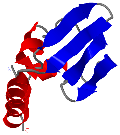
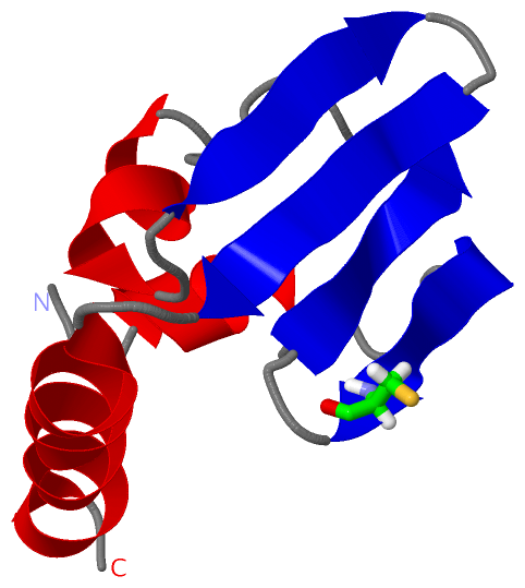
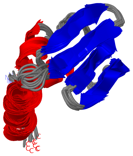
 Visualization

(-) Interactive Views

NMR Structure
  Complete Structure
    Jena3D(integrated viewing of ligand, site, SAP, PROSITE, SCOP information)
    WebMol | AstexViewer[tm]@PDBe
(Java Applets, require no local installation except for Java; loading may be slow)
    STRAP
(Java WebStart application, automatic local installation, requires Java; full application with system access!)
    RasMol
(require local installation)
    Molscript (VRML)
(requires installation of a VRML viewer; select preferred view via VRML and generate a mono or stereo PDF format file)
 
  Ligands, Modified Residues, Ions
(no "Ligands, Modified Residues, Ions" information available for 1iba)
 
  Sites
    S1  [ RasMol ]  +environment [ RasMol ]
 
  Cis Peptide Bonds
(no "Cis Peptide Bonds" information available for 1iba)
 

(-) Still Images

Jmol
  protein: cartoon or spacefill or dots and stick; nucleic acid: cartoon and stick; ligands: spacefill; active site: stick
Molscript
  protein, nucleic acid: cartoon; ligands: spacefill; active site: ball and stick

 Databases and Analysis Tools

(-) Databases

Access by PDB/NDB ID
  1iba
    Family and Domain InformationProDom | SYSTERS
    General Structural InformationGlycoscienceDB | MMDB | NDB | OCA | PDB | PDBe | PDBj | PDBsum | PDBWiki | PQS | PROTEOPEDIA
    Orientation in MembranesOPM
    Protein SurfaceSURFACE
    Secondary StructureDSSP (structure derived) | HSSP (homology derived)
    Structural GenomicsGeneCensus
    Structural NeighboursCE | VAST
    Structure ClassificationCATH | Dali | SCOP
    Validation and Original DataBMRB Data View | BMRB Restraints Grid | EDS | PROCHECK | RECOORD | WHAT_CHECK
 
Access by UniProt ID/Accession number
  PTGCB_ECOLI | P69786
    Comparative Protein Structure ModelsModBase
    Genomic InformationEnsembl
    Protein-protein InteractionDIP
    Sequence, Family and Domain InformationInterPro | Pfam | SMART | UniProtKB/SwissProt
 
Access by Enzyme Classificator   (EC Number)
  2.7.1.69
    General Enzyme InformationBRENDA | EC-PDB | Enzyme | IntEnz
    PathwayKEGG | MetaCyc
 
Access by Disease Identifier   (MIM ID)
  (no 'MIM ID' available)
    Disease InformationOMIM
 
Access by GenAge ID
  (no 'GenAge ID' available)
    Age Related InformationGenAge

(-) Analysis Tools

Access by PDB/NDB ID
    Domain InformationXDom
    Interatomic Contacts of Structural UnitsCSU
    Ligand-protein ContactsLPC
    Protein CavitiescastP
    Sequence and Secondary StructurePDBCartoon
    Structure AlignmentSTRAP(Java WebStart application, automatic local installation, requires Java; full application with system access!)
    Structure and Sequence BrowserSTING
 
Access by UniProt ID/Accession number
  PTGCB_ECOLI | P69786
    Protein Disorder PredictionDisEMBL | FoldIndex | GLOBPLOT (for more information see DisProt)

 Related Entries

(-) Entries Sharing at Least One Protein Chain (UniProt ID)

UniProtKB/Swiss-Prot
        PTGCB_ECOLI | P697861o2f 3bp3 3bp8

(-) Related Entries Specified in the PDB File

(no "Related Entries Specified in the PDB File" available for 1IBA)