|
|
|
|
 Description
|
|
Compounds
|
||||||||||||||||||||||||||||||||||||||||||||||||||||
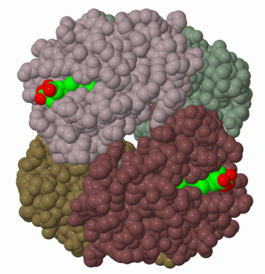
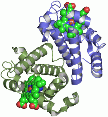
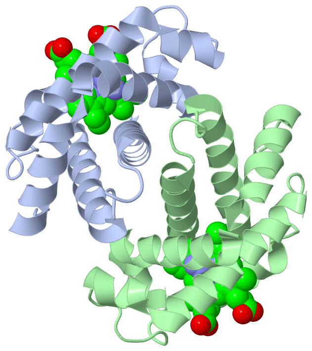
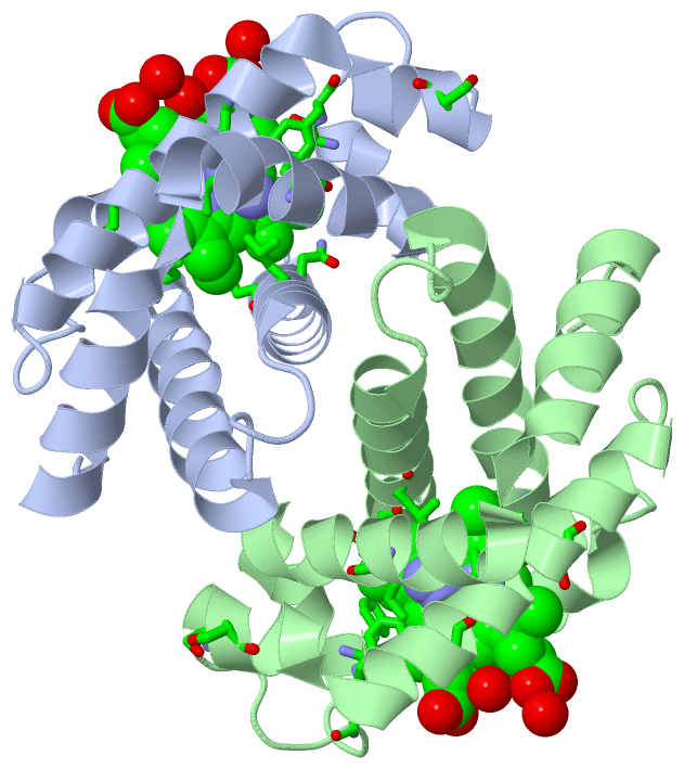
Chains, Units
Summary Information (see also Sequences/Alignments below) |
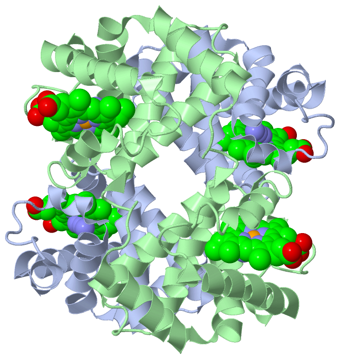
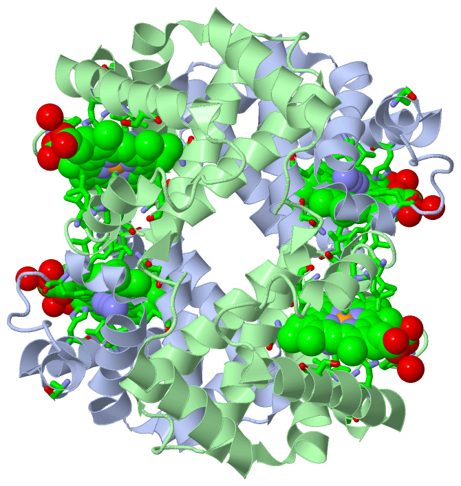
Ligands, Modified Residues, Ions (2, 4)| Asymmetric Unit (2, 4) Biological Unit 1 (2, 8) |
Sites (4, 4)
Asymmetric Unit (4, 4)
|
SS Bonds (0, 0)| (no "SS Bond" information available for 1I3E) |
Cis Peptide Bonds (0, 0)| (no "Cis Peptide Bond" information available for 1I3E) |
SAPs(SNPs)/Variants (25, 50)
Asymmetric Unit (25, 50)
|
||||||||||||||||||||||||||||||||||||||||||||||||||||||||||||||||||||||||||||||||||||||||||||||||||||||||||||||||||||||||||||||||||||||||||||||||||||||||||||||||||||||||||||||||||||||||||||||||||||||||||||||||||||||||||||||||||||||||||||||||||||||||||||||||||||||||||||||||||||||||||||||||||||||||||||||||||||||||||||||||||||||||||||||||||||||||||||||||||||||||||||||||||||||||||||||||||||||||||||||||||||||||||||||||||||||||||||||||||||||||||||||||||||||||||||||||||||||||||||||||||||||||||||||||||||||||||||||||||||||||||||||||||||||||||||||||||||||||||||||||||||||||||||||||||||||||||||||||||||||||||||||||||||||||||||||||||||||||||||||||||||||||||||||||||||||||||||||||||||||||||||||||||||||||||||||||||||||||||||||||||||||||||
PROSITE Motifs (1, 2)
Asymmetric Unit (1, 2)
|
||||||||||||||||||||||||||||||||||||||||||||||||
Exons (0, 0)| (no "Exon" information available for 1I3E) |
Sequences/Alignments
Asymmetric UnitChain A from PDB Type:PROTEIN Length:146 aligned with HBG1_HUMAN | P69891 from UniProtKB/Swiss-Prot Length:147 Alignment length:146 11 21 31 41 51 61 71 81 91 101 111 121 131 141 HBG1_HUMAN 2 GHFTEEDKATITSLWGKVNVEDAGGETLGRLLVVYPWTQRFFDSFGNLSSASAIMGNPKVKAHGKKVLTSLGDAIKHLDDLKGTFAQLSELHCDKLHVDPENFKLLGNVLVTVLAIHFGKEFTPEVQASWQKMVTAVASALSSRYH 147 SCOP domains d1i3ea_ A: Hemoglobin, beta-chain SCOP domains CATH domains 1i3eA00 A:1-146 Globins CATH domains Pfam domains -------------------------------------------------------------------------------------------------------------------------------------------------- Pfam domains SAPs(SNPs) (1) -Q--KG-----R---------G--R----------RG-RK--N---------D-------E----------RH-T---NN----------------R-----------------------K------T-----M------------ SAPs(SNPs) (1) SAPs(SNPs) (2) -----Q------------------------------------------------------------------N------Y------------------------------------------------------------------ SAPs(SNPs) (2) PROSITE GLOBIN PDB: A:1-146 UniProt: 2-147 PROSITE Transcript -------------------------------------------------------------------------------------------------------------------------------------------------- Transcript 1i3e A 1 GHFTEEDKATITSLWGKVNVEDAGGETLGRLLVVYPWTQRFFDSFGNLSSASAIMGNPKVKAHGKKVLTSLGDAIKHLDDLKGTFAQLSELHCDKLHVDPENFKLLGNVLVTVLAIHFGKEFTPEVQASWQKMVTAVASALSSRYH 146 10 20 30 40 50 60 70 80 90 100 110 120 130 140 Chain B from PDB Type:PROTEIN Length:146 aligned with HBG1_HUMAN | P69891 from UniProtKB/Swiss-Prot Length:147 Alignment length:146 11 21 31 41 51 61 71 81 91 101 111 121 131 141 HBG1_HUMAN 2 GHFTEEDKATITSLWGKVNVEDAGGETLGRLLVVYPWTQRFFDSFGNLSSASAIMGNPKVKAHGKKVLTSLGDAIKHLDDLKGTFAQLSELHCDKLHVDPENFKLLGNVLVTVLAIHFGKEFTPEVQASWQKMVTAVASALSSRYH 147 SCOP domains d1i3eb_ B: Hemoglobin, beta-chain SCOP domains CATH domains 1i3eB00 B:1-146 Globins CATH domains Pfam domains -------------------------------------------------------------------------------------------------------------------------------------------------- Pfam domains SAPs(SNPs) (1) -Q--KG-----R---------G--R----------RG-RK--N---------D-------E----------RH-T---NN----------------R-----------------------K------T-----M------------ SAPs(SNPs) (1) SAPs(SNPs) (2) -----Q------------------------------------------------------------------N------Y------------------------------------------------------------------ SAPs(SNPs) (2) PROSITE GLOBIN PDB: B:1-146 UniProt: 2-147 PROSITE Transcript -------------------------------------------------------------------------------------------------------------------------------------------------- Transcript 1i3e B 1 GHFTEEDKATITSLWGKVNVEDAGGETLGRLLVVYPWTQRFFDSFGNLSSASAIMGNPKVKAHGKKVLTSLGDAIKHLDDLKGTFAQLSELHCDKLHVDPENFKLLGNVLVTVLAIHFGKEFTPEVQASWQKMVTAVASALSSRYH 146 10 20 30 40 50 60 70 80 90 100 110 120 130 140
|
||||||||||||||||||||
SCOP Domains (1, 2)
Asymmetric Unit
 |
CATH Domains (1, 2)
Asymmetric Unit
|
Pfam Domains (0, 0)| (no "Pfam Domain" information available for 1I3E) |
Gene Ontology (10, 10)|
Asymmetric Unit(hide GO term definitions) Chain A,B (HBG1_HUMAN | P69891)
|
||||||||||||||||||||||||||||||||||||||||||||||||||||||||||||||||||||||||||||||
Interactive Views
|
||||||||||||||||||||||||||||||||||||||||||||||||||||||||||||||||||||||||||||||||||||||||||||||||||||||||||||||||||||||||||||||||||||||||||||||||||||||||||||||||||||
Still Images
|
||||||||||||||||
Databases
|
||||||||||||||||||||||||||||||||||||||||||||||||||||||||||||||||||||||||||||||||||||||||||||||||||||||||||||||||||||||||||||||||||||||||||||||||||||||||||||||||
Analysis Tools
|
|||||||||||||||||||||||||||||||||||||||||||||||||||||||||||||
Entries Sharing at Least One Protein Chain (UniProt ID)
Related Entries Specified in the PDB File
|
|