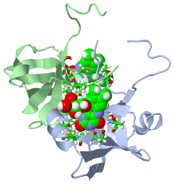
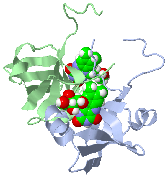
Chain A from PDB  Type:PROTEIN  Length:97
 aligned with RISA_ECOLI | P0AFU8 from UniProtKB/Swiss-Prot  Length:213

    Alignment length:97
                                    10        20        30        40        50        60        70        80        90
            RISA_ECOLI    1 MFTGIVQGTAKLVSIDEKPNFRTHVVELPDHMLDGLETGASVAHNGCCLTVTEINGNHVSFDLMKETLRITNLGDLKVGDWVNVERAAKFSDEIGGH 97
               SCOP domains d1hzea_ A: Riboflavin synthase                                                                    SCOP domains
               CATH domains 1hzeA00 A:1-97  [code=2.40.30.20, no name defined]                                                CATH domains
               Pfam domains ------------------------------------------------------------------------------------------------- Pfam domains
         Sec.struct. author .......eeeeeeeeee....eeeeee.hhhhhh......eeee..eeeeeeeee..eeeeeehhhhhhhhhhhhh....eeeeeee.......... Sec.struct. author
                 SAPs(SNPs) ------------------------------------------------------------------------------------------------- SAPs(SNPs)
                PROSITE (2) LUMAZINE_BIND  PDB: A:1-97 UniProt: 1-97                                                          PROSITE (2)
                 Transcript ------------------------------------------------------------------------------------------------- Transcript
                  1hze A  1 MFTGIVQGTAKLVSIDEKPNFRTHVVELPDHMLDGLETGASVAHNGCCLTVTEINGNHVSFDLMKETLRITNLGDLKVGDWVNVERAAKFSDEIGGH 97
                                    10        20        30        40        50        60        70        80        90
Chain A from PDB  Type:PROTEIN  Length:97
 aligned with RISA_SHIFL | P0AFU9 from UniProtKB/Swiss-Prot  Length:213

    Alignment length:97
                                    10        20        30        40        50        60        70        80        90
            RISA_SHIFL    1 MFTGIVQGTAKLVSIDEKPNFRTHVVELPDHMLDGLETGASVAHNGCCLTVTEINGNHVSFDLMKETLRITNLGDLKVGDWVNVERAAKFSDEIGGH 97
               SCOP domains d1hzea_ A: Riboflavin synthase                                                                    SCOP domains
               CATH domains 1hzeA00 A:1-97  [code=2.40.30.20, no name defined]                                                CATH domains
               Pfam domains ------------------------------------------------------------------------------------------------- Pfam domains
         Sec.struct. author .......eeeeeeeeee....eeeeee.hhhhhh......eeee..eeeeeeeee..eeeeeehhhhhhhhhhhhh....eeeeeee.......... Sec.struct. author
                 SAPs(SNPs) ------------------------------------------------------------------------------------------------- SAPs(SNPs)
                    PROSITE LUMAZINE_BIND  PDB: A:1-97 UniProt: 1-97                                                          PROSITE
                 Transcript ------------------------------------------------------------------------------------------------- Transcript
                  1hze A  1 MFTGIVQGTAKLVSIDEKPNFRTHVVELPDHMLDGLETGASVAHNGCCLTVTEINGNHVSFDLMKETLRITNLGDLKVGDWVNVERAAKFSDEIGGH 97
                                    10        20        30        40        50        60        70        80        90
Chain B from PDB  Type:PROTEIN  Length:97
 aligned with RISA_ECOLI | P0AFU8 from UniProtKB/Swiss-Prot  Length:213

    Alignment length:97
                                    10        20        30        40        50        60        70        80        90
            RISA_ECOLI    1 MFTGIVQGTAKLVSIDEKPNFRTHVVELPDHMLDGLETGASVAHNGCCLTVTEINGNHVSFDLMKETLRITNLGDLKVGDWVNVERAAKFSDEIGGH 97
               SCOP domains d1hzeb_ B: Riboflavin synthase                                                                    SCOP domains
               CATH domains 1hzeB00 B:1-97  [code=2.40.30.20, no name defined]                                                CATH domains
               Pfam domains ------------------------------------------------------------------------------------------------- Pfam domains
         Sec.struct. author .......eeeeeeeeee....eeeeee.hhhhhh......eeee..eeeeeeeee..eeeeeehhhhhhhhhhhhh....eeeeeee.......... Sec.struct. author
                 SAPs(SNPs) ------------------------------------------------------------------------------------------------- SAPs(SNPs)
                PROSITE (2) LUMAZINE_BIND  PDB: B:1-97 UniProt: 1-97                                                          PROSITE (2)
                 Transcript ------------------------------------------------------------------------------------------------- Transcript
                  1hze B  1 MFTGIVQGTAKLVSIDEKPNFRTHVVELPDHMLDGLETGASVAHNGCCLTVTEINGNHVSFDLMKETLRITNLGDLKVGDWVNVERAAKFSDEIGGH 97
                                    10        20        30        40        50        60        70        80        90
Chain B from PDB  Type:PROTEIN  Length:97
 aligned with RISA_SHIFL | P0AFU9 from UniProtKB/Swiss-Prot  Length:213

    Alignment length:97
                                    10        20        30        40        50        60        70        80        90
            RISA_SHIFL    1 MFTGIVQGTAKLVSIDEKPNFRTHVVELPDHMLDGLETGASVAHNGCCLTVTEINGNHVSFDLMKETLRITNLGDLKVGDWVNVERAAKFSDEIGGH 97
               SCOP domains d1hzeb_ B: Riboflavin synthase                                                                    SCOP domains
               CATH domains 1hzeB00 B:1-97  [code=2.40.30.20, no name defined]                                                CATH domains
               Pfam domains ------------------------------------------------------------------------------------------------- Pfam domains
         Sec.struct. author .......eeeeeeeeee....eeeeee.hhhhhh......eeee..eeeeeeeee..eeeeeehhhhhhhhhhhhh....eeeeeee.......... Sec.struct. author
                 SAPs(SNPs) ------------------------------------------------------------------------------------------------- SAPs(SNPs)
                    PROSITE LUMAZINE_BIND  PDB: B:1-97 UniProt: 1-97                                                          PROSITE
                 Transcript ------------------------------------------------------------------------------------------------- Transcript
                  1hze B  1 MFTGIVQGTAKLVSIDEKPNFRTHVVELPDHMLDGLETGASVAHNGCCLTVTEINGNHVSFDLMKETLRITNLGDLKVGDWVNVERAAKFSDEIGGH 97
                                    10        20        30        40        50        60        70        80        90
| Legend: |
|
→ Mismatch |
(orange background) |
| |
- |
→ Gap |
(green background, '-', border residues have a numbering label) |
| |
|
→ Modified Residue |
(blue background, lower-case, 'x' indicates undefined single-letter code, labelled with number + name) |
| |
x |
→ Chemical Group |
(purple background, 'x', labelled with number + name, e.g. ACE or NH2) |
| |
extra numbering lines below/above indicate numbering irregularities and modified residue names etc., number ends below/above '|' |
 Description