|
|
|
|
 Description
|
|
Compounds
|
||||||||||||||||||||||||||||||||||||||||||||||||||||
Chains, Units
Summary Information (see also Sequences/Alignments below) |
Ligands, Modified Residues, Ions (0, 0)| (no "Ligand,Modified Residues,Ions" information available for 1HPW) |
Sites (0, 0)| (no "Site" information available for 1HPW) |
SS Bonds (2, 2)
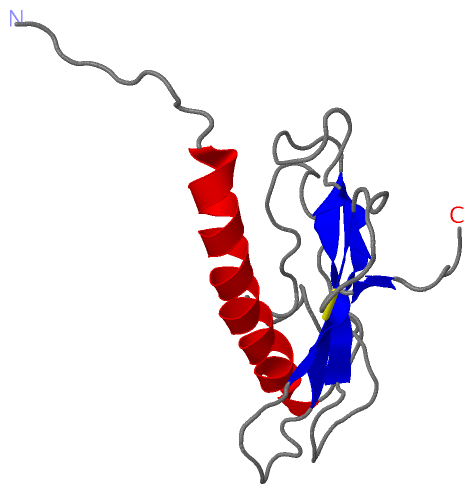
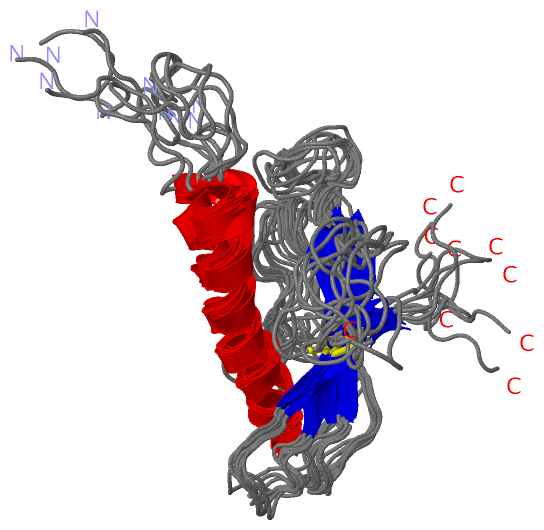
NMR Structure
|
||||||||||||
Cis Peptide Bonds (0, 0)| (no "Cis Peptide Bond" information available for 1HPW) |
SAPs(SNPs)/Variants (0, 0)| (no "SAP(SNP)/Variant" information available for 1HPW) |
PROSITE Motifs (1, 1)
NMR Structure (1, 1)
|
||||||||||||||||||||||||
Exons (0, 0)| (no "Exon" information available for 1HPW) |
Sequences/Alignments
NMR StructureChain A from PDB Type:PROTEIN Length:129 aligned with FMP1_PSEAI | P17838 from UniProtKB/Swiss-Prot Length:157 Alignment length:154 13 23 33 43 53 63 73 83 93 103 113 123 133 143 153 FMP1_PSEAI 4 AQKGFTLIELMIVVAIIGILAAIAIPAYQDYTARAQLSERMTLASGLKTKVSDIFSQDGSCPANTAATAGIEKDTDINGKYVAKVTTGGTAAASGGCTIVATMKASDVATPLRGKTLTLTLGNADKGSYTWACTSNADNKYLPKTCQTATTTTP 157 SCOP domains d1hp wa_ A: Pilin P1 SCOP domains CATH domains 1hpw A00 A:22-150 Glycoprotein, Type 4 Pilin CATH domains Pfam domains ---------------------------------------------------------------------------------------------------------------------------------------------------------- Pfam domains SAPs(SNPs) ---------------------------------------------------------------------------------------------------------------------------------------------------------- SAPs(SNPs) PROSITE --PROKAR_NTER_METHYL ----------------------------------------------------------------------------------------------------------------------------------- PROSITE Transcript ---------------------------------------------------------------------------------------------------------------------------------------------------------- Transcript 1hpw A 22 ALEG------------------------TEF-ARAQLSEAMTLASGLKTKVSDIFSQDGSCPANTAATAGIEKDTDINGKYVAKVTTGGTAAASGGCTIVATMKASDVATPLRGKTLTLTLGNADKGSYTWACTSNADNKYLPKTCQTATTTTP 150 | - - 27| | 36 46 56 66 76 86 96 106 116 126 136 146 25 26 | | 28 | 29
|
||||||||||||||||||||
SCOP Domains (1, 1)
NMR Structure
 |
CATH Domains (1, 1)
NMR Structure
|
Pfam Domains (0, 0)| (no "Pfam Domain" information available for 1HPW) |
Gene Ontology (2, 2)|
NMR Structure(hide GO term definitions) Chain A (FMP1_PSEAI | P17838)
|
||||||||||||||||||||||||
Interactive Views
|
||||||||||||||||||||||||||||||||||||||||||||||||||||||||||||||||||||||||||||||||||||||||||||||||||||||||||||||||||||
Still Images
|
||||||||||||||||
Databases
|
||||||||||||||||||||||||||||||||||||||||||||||||||||||||||||||||||||||||||||||||||||||||||||||||||||||||||||||||||||||||||||||||||||||||||||||||||||||||||||||||
Analysis Tools
|
|||||||||||||||||||||||||||||||||||||||||||||||||||||||||||||
Entries Sharing at Least One Protein Chain (UniProt ID)
Related Entries Specified in the PDB File
|
|