|
|
|
|
 Description
|
|
Compounds
|
||||||||||||||||||||
Chains, Units
Summary Information (see also Sequences/Alignments below) |
Ligands, Modified Residues, Ions (0, 0)| (no "Ligand,Modified Residues,Ions" information available for 1HNR) |
Sites (0, 0)| (no "Site" information available for 1HNR) |
SS Bonds (0, 0)| (no "SS Bond" information available for 1HNR) |
Cis Peptide Bonds (0, 0)| (no "Cis Peptide Bond" information available for 1HNR) |
SAPs(SNPs)/Variants (0, 0)| (no "SAP(SNP)/Variant" information available for 1HNR) |
PROSITE Motifs (0, 0)| (no "PROSITE Motif" information available for 1HNR) |
Exons (0, 0)| (no "Exon" information available for 1HNR) |
Sequences/Alignments
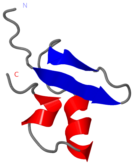
NMR StructureChain A from PDB Type:PROTEIN Length:47 aligned with HNS_ECOLI | P0ACF8 from UniProtKB/Swiss-Prot Length:137 Alignment length:47 100 110 120 130 HNS_ECOLI 91 AQRPAKYSYVDENGETKTWTGQGRTPAVIKKAMDEQGKSLDDFLIKQ 137 SCOP domains d1hnra_ A: H1 protein (H-NS) SCOP domains CATH domains 1hnrA00 A:90-136 H-NS DNA Binding Protein CATH domains Pfam domains ----------------------------------------------- Pfam domains SAPs(SNPs) ----------------------------------------------- SAPs(SNPs) PROSITE ----------------------------------------------- PROSITE Transcript ----------------------------------------------- Transcript 1hnr A 90 AQRPAKYSYVDENGETKTWTGQGRTPAVIKKAMDEQGKSLDDFLIKQ 136 99 109 119 129
|
||||||||||||||||||||
SCOP Domains (1, 1)
NMR Structure
 |
CATH Domains (1, 1)
NMR Structure
|
Pfam Domains (0, 0)| (no "Pfam Domain" information available for 1HNR) |
Gene Ontology (10, 10)|
NMR Structure(hide GO term definitions) Chain A (HNS_ECOLI | P0ACF8)
|
||||||||||||||||||||||||||||||||||||||||||||||||||||||||||||||||||||||||||||||
Interactive Views
|
||||||||||||||||||||||||||||||||||||||||||||||||||||||||||||||||||||||||||||||||||||||||||||||||||||||||||||||||||||
Still Images
|
||||||||||||||||
Databases
|
||||||||||||||||||||||||||||||||||||||||||||||||||||||||||||||||||||||||||||||||||||||||||||||||||||||||||||||||||||||||||||||||||||||||||||||||||||||||||||||||
Analysis Tools
|
|||||||||||||||||||||||||||||||||||||||||||||||||||||||||||||
Entries Sharing at Least One Protein Chain (UniProt ID)
Related Entries Specified in the PDB File
|
|