Show PDB file:   
         Plain Text   HTML   (compressed file size)
QuickSearch:   
by PDB,NDB,UniProt,PROSITE Code or Search Term(s)  
(-)Asymmetric Unit
(-)Asym. Unit - sites
(-)Biological Unit 1
(-)Biol. Unit 1 - sites
(-)Biological Unit 2
collapse expand < >
Image Asymmetric Unit
Asymmetric Unit  (Jmol Viewer)
Image Asym. Unit - sites
Asym. Unit - sites  (Jmol Viewer)
Image Biological Unit 1
Biological Unit 1  (Jmol Viewer)
Image Biol. Unit 1 - sites
Biol. Unit 1 - sites  (Jmol Viewer)
Image Biological Unit 2
Biological Unit 2  (Jmol Viewer)

(-) Description

Title :  DECAPLANIN FIRST P21-FORM
 
Authors :  C. Lehmann, L. Vertesy, G. M. Sheldrick, Z. Dauter, M. Dauter
Date :  19 Dec 00  (Deposition) - 11 Jul 05  (Release) - 01 May 13  (Revision)
Method :  X-RAY DIFFRACTION
Resolution :  1.00
Chains :  Asym. Unit :  A,B,C,D
Biol. Unit 1:  A,B  (1x)
Biol. Unit 2:  C,D  (1x)
Keywords :  Antibiotic, Glycopeptide (Keyword Search: [Gene Ontology, PubMed, Web (Google))
 
Reference :  C. Lehmann, J. E. Debreczeni, G. Bunkoczi, M. Dauter, Z. Dauter, L. Vertesy, G. M. Sheldrick
Structures Of Four Crystal Forms Of Decaplanin
Helv. Chim. Acta V. 86 1478 2003
PubMed: search  |  Reference-DOI: 10.1002/HLCA.200390131

(-) Compounds

Molecule 1 - DECAPLANIN
    ChainsA, B, C, D
    Organism ScientificUNCULTURED ACTINOMYCETE
    Organism Taxid100235
    Other DetailsCULTURE Y-86, 36910
    StrainDSM 4763
    SynonymM86-1410

 Structural Features

(-) Chains, Units

  1234
Asymmetric Unit ABCD
Biological Unit 1 (1x)AB  
Biological Unit 2 (1x)  CD

Summary Information (see also Sequences/Alignments below)

(-) Ligands, Modified Residues, Ions  (9, 39)

Asymmetric Unit (9, 39)
No.NameCountTypeFull Name
13FG4Mod. Amino Acid(2S)-AMINO(3,5-DIHYDROXYPHENYL)ETHANOIC ACID
2BGC4Ligand/IonBETA-D-GLUCOSE
3ERE4Ligand/Ion(1R,3S,4R,5S)-3-AMINO-2,3,6-TRIDEOXY-3-METHYL-ALPHA-L-ARABINO-HEXOPYRANOSE
4GHP8Mod. Amino Acid(2R)-AMINO(4-HYDROXYPHENYL)ETHANOIC ACID
5GOL3Ligand/IonGLYCEROL
6MLU4Mod. Amino AcidN-METHYL-D-LEUCINE
7OMX4Mod. Amino Acid(BETAR)-BETA-HYDROXY-L-TYROSINE
8OMZ4Mod. Amino Acid(BETAR)-3-CHLORO-BETA-HYDROXY-D-TYROSINE
9RAM4Ligand/IonALPHA-L-RHAMNOSE
Biological Unit 1 (9, 20)
No.NameCountTypeFull Name
13FG2Mod. Amino Acid(2S)-AMINO(3,5-DIHYDROXYPHENYL)ETHANOIC ACID
2BGC2Ligand/IonBETA-D-GLUCOSE
3ERE2Ligand/Ion(1R,3S,4R,5S)-3-AMINO-2,3,6-TRIDEOXY-3-METHYL-ALPHA-L-ARABINO-HEXOPYRANOSE
4GHP4Mod. Amino Acid(2R)-AMINO(4-HYDROXYPHENYL)ETHANOIC ACID
5GOL2Ligand/IonGLYCEROL
6MLU2Mod. Amino AcidN-METHYL-D-LEUCINE
7OMX2Mod. Amino Acid(BETAR)-BETA-HYDROXY-L-TYROSINE
8OMZ2Mod. Amino Acid(BETAR)-3-CHLORO-BETA-HYDROXY-D-TYROSINE
9RAM2Ligand/IonALPHA-L-RHAMNOSE
Biological Unit 2 (9, 19)
No.NameCountTypeFull Name
13FG2Mod. Amino Acid(2S)-AMINO(3,5-DIHYDROXYPHENYL)ETHANOIC ACID
2BGC2Ligand/IonBETA-D-GLUCOSE
3ERE2Ligand/Ion(1R,3S,4R,5S)-3-AMINO-2,3,6-TRIDEOXY-3-METHYL-ALPHA-L-ARABINO-HEXOPYRANOSE
4GHP4Mod. Amino Acid(2R)-AMINO(4-HYDROXYPHENYL)ETHANOIC ACID
5GOL1Ligand/IonGLYCEROL
6MLU2Mod. Amino AcidN-METHYL-D-LEUCINE
7OMX2Mod. Amino Acid(BETAR)-BETA-HYDROXY-L-TYROSINE
8OMZ2Mod. Amino Acid(BETAR)-3-CHLORO-BETA-HYDROXY-D-TYROSINE
9RAM2Ligand/IonALPHA-L-RHAMNOSE

(-) Sites  (7, 7)

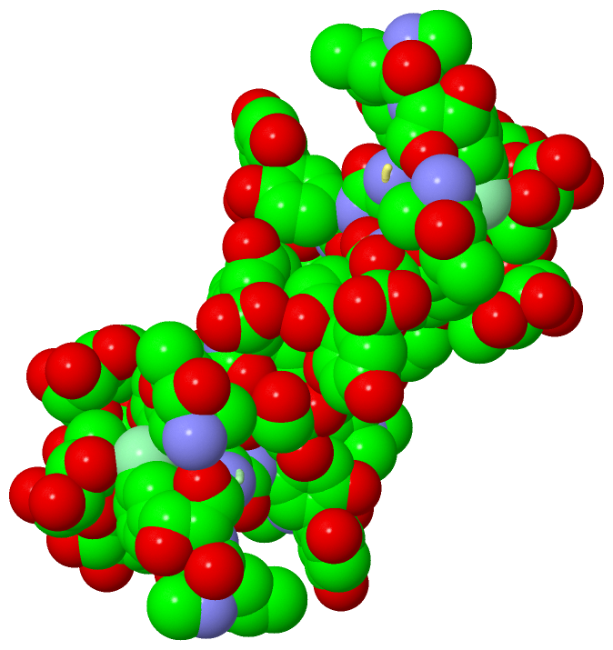
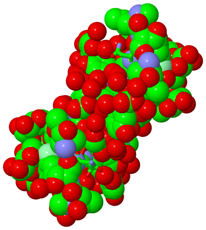
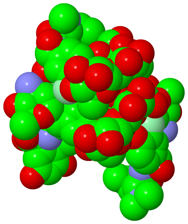
Asymmetric Unit (7, 7)
No.NameEvidenceResiduesDescription
1AC1SOFTWAREMLU B:1 , OMZ B:2 , GHP B:4 , HOH B:2007 , HOH B:2042 , HOH B:2043 , HOH B:2044 , GHP D:5 , 3FG D:7BINDING SITE FOR RESIDUE GOL B1001
2AC2SOFTWAREGHP A:5 , OMX A:6 , HOH A:2024 , ASN B:3 , HOH B:2046 , HOH B:2047 , 3FG C:7 , MLU D:1BINDING SITE FOR RESIDUE GOL B1002
3AC3SOFTWAREGHP B:5 , 3FG B:7 , MLU D:1 , OMZ D:2 , HOH D:2001 , HOH D:2011 , HOH D:2032 , HOH D:2033BINDING SITE FOR RESIDUE GOL D1003
4AC4SOFTWAREHOH A:2001 , HOH A:2002 , HOH A:2009 , HOH A:2010 , HOH A:2011 , HOH A:2014 , HOH A:2023 , HOH A:2025 , HOH A:2026 , HOH A:2028 , HOH A:2029 , HOH A:2030 , HOH A:2031 , HOH A:2032 , ASN B:3 , GHP B:4 , GHP B:5 , OMX B:6 , GOL B:1002 , MLU C:1 , OMZ C:2 , ASN C:3 , GHP C:5 , GHP D:5BINDING SITE FOR CHAIN A OF DECAPLANIN
5AC5SOFTWAREASN A:3 , GHP A:4 , GHP A:5 , OMX A:6 , GOL B:1001 , GOL B:1002 , HOH B:2001 , HOH B:2002 , HOH B:2006 , HOH B:2007 , HOH B:2008 , HOH B:2011 , HOH B:2012 , HOH B:2020 , HOH B:2021 , HOH B:2022 , HOH B:2023 , HOH B:2024 , HOH B:2025 , HOH B:2026 , HOH B:2028 , MLU C:1 , MLU D:1 , ASN D:3 , GHP D:5 , OMX D:6 , 3FG D:7 , GOL D:1003 , HOH D:2006 , HOH D:2011 , HOH D:2013 , HOH D:2018 , HOH D:2032BINDING SITE FOR CHAIN B OF DECAPLANIN
6AC6SOFTWAREASN A:3 , GHP A:4 , GHP A:5 , 3FG A:7 , HOH A:2023 , HOH A:2024 , GHP B:5 , GOL B:1002 , HOH B:2020 , HOH B:2022 , HOH B:2023 , HOH C:2001 , HOH C:2009 , HOH C:2010 , HOH C:2011 , HOH C:2012 , HOH C:2022 , HOH C:2024 , HOH C:2025 , HOH C:2026 , HOH C:2027 , HOH C:2028 , HOH C:2029 , HOH C:2030 , OMZ D:2 , ASN D:3 , GHP D:4 , GHP D:5 , OMX D:6BINDING SITE FOR CHAIN C OF DECAPLANIN
7AC7SOFTWARE3FG A:7 , HOH A:2029 , HOH A:2032 , MLU B:1 , ASN B:3 , GHP B:5 , OMX B:6 , 3FG B:7 , GOL B:1001 , GOL B:1002 , HOH B:2011 , HOH B:2012 , HOH B:2042 , HOH B:2046 , OMZ C:2 , ASN C:3 , GHP C:4 , GHP C:5 , OMX C:6 , HOH C:2024 , HOH C:2032 , GOL D:1003 , HOH D:2001 , HOH D:2005 , HOH D:2006 , HOH D:2011 , HOH D:2012 , HOH D:2013 , HOH D:2014 , HOH D:2015 , HOH D:2016 , HOH D:2017 , HOH D:2018 , HOH D:2019BINDING SITE FOR CHAIN D OF DECAPLANIN

(-) SS Bonds  (0, 0)

(no "SS Bond" information available for 1HH3)

(-) Cis Peptide Bonds  (4, 4)

Asymmetric Unit
No.Residues
1Ghp A:5 -Omx A:6
2Ghp B:5 -Omx B:6
3Ghp C:5 -Omx C:6
4Ghp D:5 -Omx D:6

 Sequence-Structure Mapping

(-) SAPs(SNPs)/Variants  (0, 0)

(no "SAP(SNP)/Variant" information available for 1HH3)

(-) PROSITE Motifs  (0, 0)

(no "PROSITE Motif" information available for 1HH3)

(-) Exons   (0, 0)

(no "Exon" information available for 1HH3)

(-) Sequences/Alignments

Asymmetric Unit
   Reformat: Number of residues per line =  ('0' or empty: single-line sequence representation)
  Number of residues per labelling interval =   
  UniProt sequence: complete  aligned part    
   Show mapping: SCOP domains CATH domains Pfam domains Secondary structure (by author)
SAPs(SNPs) PROSITE motifs Exons
(details for a mapped element are shown in a popup box when the mouse pointer rests over it)
Chain A from PDB  Type:PROTEIN  Length:7
                                     
               SCOP domains ------- SCOP domains
               CATH domains ------- CATH domains
               Pfam domains ------- Pfam domains
         Sec.struct. author ....... Sec.struct. author
                 SAPs(SNPs) ------- SAPs(SNPs)
                    PROSITE ------- PROSITE
                 Transcript ------- Transcript
                   1hh3 A 1 xxNxxxx 7
                            || ||||
                            || ||||
                            1-MLU||
                             2-OMZ|
                               4-GHP
                                5-GHP
                                 6-OMX
                                  7-3FG

Chain B from PDB  Type:PROTEIN  Length:7
                                     
               SCOP domains ------- SCOP domains
               CATH domains ------- CATH domains
               Pfam domains ------- Pfam domains
         Sec.struct. author ....... Sec.struct. author
                 SAPs(SNPs) ------- SAPs(SNPs)
                    PROSITE ------- PROSITE
                 Transcript ------- Transcript
                   1hh3 B 1 xxNxxxx 7
                            || ||||
                            || ||||
                            1-MLU||
                             2-OMZ|
                               4-GHP
                                5-GHP
                                 6-OMX
                                  7-3FG

Chain C from PDB  Type:PROTEIN  Length:7
                                     
               SCOP domains ------- SCOP domains
               CATH domains ------- CATH domains
               Pfam domains ------- Pfam domains
         Sec.struct. author ....... Sec.struct. author
                 SAPs(SNPs) ------- SAPs(SNPs)
                    PROSITE ------- PROSITE
                 Transcript ------- Transcript
                   1hh3 C 1 xxNxxxx 7
                            || ||||
                            || ||||
                            1-MLU||
                             2-OMZ|
                               4-GHP
                                5-GHP
                                 6-OMX
                                  7-3FG

Chain D from PDB  Type:PROTEIN  Length:7
                                     
               SCOP domains ------- SCOP domains
               CATH domains ------- CATH domains
               Pfam domains ------- Pfam domains
         Sec.struct. author ....... Sec.struct. author
                 SAPs(SNPs) ------- SAPs(SNPs)
                    PROSITE ------- PROSITE
                 Transcript ------- Transcript
                   1hh3 D 1 xxNxxxx 7
                            || ||||
                            || ||||
                            1-MLU||
                             2-OMZ|
                               4-GHP
                                5-GHP
                                 6-OMX
                                  7-3FG

   Legend:   → Mismatch (orange background)
  - → Gap (green background, '-', border residues have a numbering label)
    → Modified Residue (blue background, lower-case, 'x' indicates undefined single-letter code, labelled with number + name)
  x → Chemical Group (purple background, 'x', labelled with number + name, e.g. ACE or NH2)
  extra numbering lines below/above indicate numbering irregularities and modified residue names etc., number ends below/above '|'

 Classification and Annotation

(-) SCOP Domains  (0, 0)

(no "SCOP Domain" information available for 1HH3)

(-) CATH Domains  (0, 0)

(no "CATH Domain" information available for 1HH3)

(-) Pfam Domains  (0, 0)

(no "Pfam Domain" information available for 1HH3)

(-) Gene Ontology  (0, 0)

Asymmetric Unit(hide GO term definitions)
    (no "Gene Ontology" information available for 1HH3)

 Visualization

(-) Interactive Views

Asymmetric Unit
  Complete Structure
    Jena3D(integrated viewing of ligand, site, SAP, PROSITE, SCOP information)
    WebMol | AstexViewer[tm]@PDBe
(Java Applets, require no local installation except for Java; loading may be slow)
    STRAP
(Java WebStart application, automatic local installation, requires Java; full application with system access!)
    RasMol
(require local installation)
    Molscript (VRML)
(requires installation of a VRML viewer; select preferred view via VRML and generate a mono or stereo PDF format file)
 
  Ligands, Modified Residues, Ions
    3FG  [ RasMol | Jena3D ]  +environment [ RasMol | Jena3D ]
    BGC  [ RasMol | Jena3D ]  +environment [ RasMol | Jena3D ]
    ERE  [ RasMol | Jena3D ]  +environment [ RasMol | Jena3D ]
    GHP  [ RasMol | Jena3D ]  +environment [ RasMol | Jena3D ]
    GOL  [ RasMol | Jena3D ]  +environment [ RasMol | Jena3D ]
    MLU  [ RasMol | Jena3D ]  +environment [ RasMol | Jena3D ]
    OMX  [ RasMol | Jena3D ]  +environment [ RasMol | Jena3D ]
    OMZ  [ RasMol | Jena3D ]  +environment [ RasMol | Jena3D ]
    RAM  [ RasMol | Jena3D ]  +environment [ RasMol | Jena3D ]
 
  Sites
    AC1  [ RasMol ]  +environment [ RasMol ]
    AC2  [ RasMol ]  +environment [ RasMol ]
    AC3  [ RasMol ]  +environment [ RasMol ]
    AC4  [ RasMol ]  +environment [ RasMol ]
    AC5  [ RasMol ]  +environment [ RasMol ]
    AC6  [ RasMol ]  +environment [ RasMol ]
    AC7  [ RasMol ]  +environment [ RasMol ]
 
  Cis Peptide Bonds
    Ghp A:5 - Omx A:6   [ RasMol ]  
    Ghp B:5 - Omx B:6   [ RasMol ]  
    Ghp C:5 - Omx C:6   [ RasMol ]  
    Ghp D:5 - Omx D:6   [ RasMol ]  
 
Biological Units
  Complete Structure
    Biological Unit 1  [ Jena3D ]
    Biological Unit 2  [ Jena3D ]

(-) Still Images

Jmol
  protein: cartoon or spacefill or dots and stick; nucleic acid: cartoon and stick; ligands: spacefill; active site: stick
Molscript
  protein, nucleic acid: cartoon; ligands: spacefill; active site: ball and stick

 Databases and Analysis Tools

(-) Databases

Access by PDB/NDB ID
  1hh3
    Family and Domain InformationProDom | SYSTERS
    General Structural InformationGlycoscienceDB | MMDB | NDB | OCA | PDB | PDBe | PDBj | PDBsum | PDBWiki | PQS | PROTEOPEDIA
    Orientation in MembranesOPM
    Protein SurfaceSURFACE
    Secondary StructureDSSP (structure derived) | HSSP (homology derived)
    Structural GenomicsGeneCensus
    Structural NeighboursCE | VAST
    Structure ClassificationCATH | Dali | SCOP
    Validation and Original DataBMRB Data View | BMRB Restraints Grid | EDS | PROCHECK | RECOORD | WHAT_CHECK
 
Access by UniProt ID/Accession number
  (no 'UniProt ID/Accession number' available) |
    Comparative Protein Structure ModelsModBase
    Genomic InformationEnsembl
    Protein-protein InteractionDIP
    Sequence, Family and Domain InformationInterPro | Pfam | SMART | UniProt
 
Access by Enzyme Classificator   (EC Number)
  (no 'Enzyme Classificator' available)
    General Enzyme InformationBRENDA | EC-PDB | Enzyme | IntEnz
    PathwayKEGG | MetaCyc
 
Access by Disease Identifier   (MIM ID)
  (no 'MIM ID' available)
    Disease InformationOMIM
 
Access by GenAge ID
  (no 'GenAge ID' available)
    Age Related InformationGenAge

(-) Analysis Tools

Access by PDB/NDB ID
    Domain InformationXDom
    Interatomic Contacts of Structural UnitsCSU
    Ligand-protein ContactsLPC
    Protein CavitiescastP
    Sequence and Secondary StructurePDBCartoon
    Structure AlignmentSTRAP(Java WebStart application, automatic local installation, requires Java; full application with system access!)
    Structure and Sequence BrowserSTING
 
Access by UniProt ID/Accession number
  (no 'UniProt ID/Accession number' available) |
    Protein Disorder PredictionDisEMBL | FoldIndex | GLOBPLOT

 Related Entries

(-) Entries Sharing at Least One Protein Chain (UniProt ID)

(no "Entries Sharing at Least One Protein Chain" available for 1HH3)

(-) Related Entries Specified in the PDB File

1hha CRYSTAL STRUCTURE OF DECAPLANIN - SPACE GROUP P6122
1hhc CRYSTAL STRUCTURE OF DECAPLANIN - SPACE GROUP P21, SECOND FORM
1hhf CRYSTAL STRUCTURE OF DECAPLANIN - SPACE GROUP P6122, SECOND FORM