Show PDB file:   
         Plain Text   HTML   (compressed file size)
QuickSearch:   
by PDB,NDB,UniProt,PROSITE Code or Search Term(s)  
(-)Asym./Biol. Unit
(-)Asym./Biol. Unit - sites
collapse expand < >
Image Asym./Biol. Unit
Asym./Biol. Unit  (Jmol Viewer)
Image Asym./Biol. Unit - sites
Asym./Biol. Unit - sites  (Jmol Viewer)

(-) Description

Title :  DTDP-MANGANESE COMPLEX OF SPSA FROM BACILLUS SUBTILIS
 
Authors :  N. Tarbouriech, S. J. Charnock, G. J. Davies
Date :  09 Jul 01  (Deposition) - 13 Dec 01  (Release) - 24 Feb 09  (Revision)
Method :  X-RAY DIFFRACTION
Resolution :  2.00
Chains :  Asym./Biol. Unit :  A
Keywords :  Transferase, Glycosyltransferase (Keyword Search: [Gene Ontology, PubMed, Web (Google))
 
Reference :  N. Tarbouriech, S. J. Charnock, G. J. Davies
Three-Dimensional Structures Of The Mn And Mg Dtdp Complexes Of The Family Gt-2 Glycosyltransferase Spsa: A Comparison With Related Ndp-Sugar Glycosyltransferases.
J. Mol. Biol. V. 314 655 2001
PubMed-ID: 11733986  |  Reference-DOI: 10.1006/JMBI.2001.5159

(-) Compounds

Molecule 1 - SPORE COAT POLYSACCHARIDE BIOSYNTHESIS PROTEIN SPSA
    ChainsA
    EngineeredYES
    Expression SystemESCHERICHIA COLI
    Expression System PlasmidPET22B
    Expression System StrainBL21
    Expression System Taxid511693
    Organism ScientificBACILLUS SUBTILIS
    Organism Taxid1423
    Other DetailsMAGNESIUM MANGANESE DTDP

 Structural Features

(-) Chains, Units

  1
Asymmetric/Biological Unit A

Summary Information (see also Sequences/Alignments below)
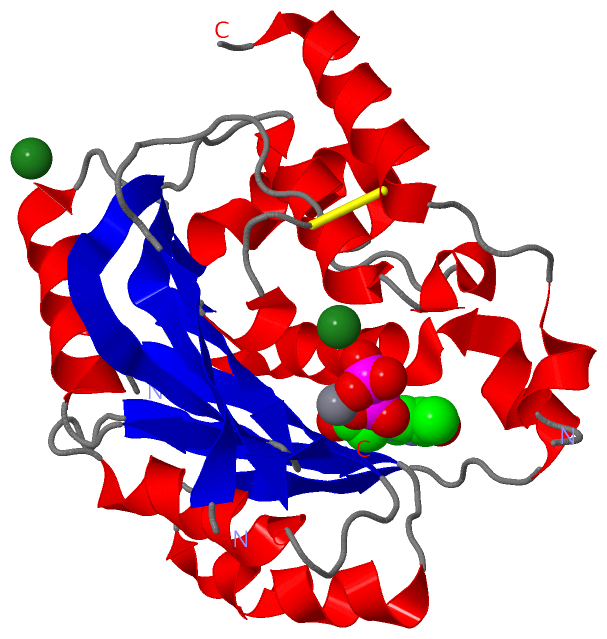
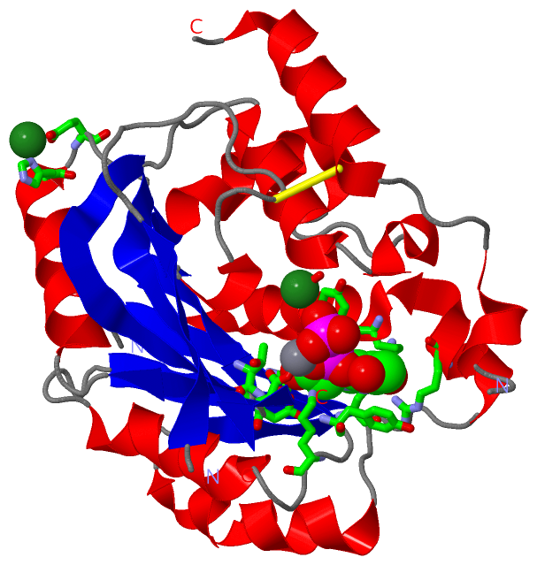
(-) Ligands, Modified Residues, Ions  (3, 4)

Asymmetric/Biological Unit (3, 4)
No.NameCountTypeFull Name
1MG2Ligand/IonMAGNESIUM ION
2MN1Ligand/IonMANGANESE (II) ION
3TYD1Ligand/IonTHYMIDINE-5'-DIPHOSPHATE

(-) Sites  (4, 4)

Asymmetric Unit (4, 4)
No.NameEvidenceResiduesDescription
1AC1SOFTWAREASP A:99 , TYD A:1258 , HOH A:2177BINDING SITE FOR RESIDUE MN A1257
2AC2SOFTWAREHIS A:117 , GLU A:119 , HOH A:2104BINDING SITE FOR RESIDUE MG A1259
3AC3SOFTWARETYD A:1258 , HOH A:2151 , HOH A:2152BINDING SITE FOR RESIDUE MG A1260
4AC4SOFTWARETHR A:9 , TYR A:11 , LYS A:13 , ASP A:39 , ARG A:71 , ARG A:76 , TYR A:77 , LEU A:80 , THR A:97 , ASP A:98 , ASP A:99 , MN A:1257 , MG A:1260 , HOH A:2177 , HOH A:2178BINDING SITE FOR RESIDUE TYD A1258

(-) SS Bonds  (1, 1)

Asymmetric/Biological Unit
No.Residues
1A:155 -A:243

(-) Cis Peptide Bonds  (0, 0)

(no "Cis Peptide Bond" information available for 1H7Q)

 Sequence-Structure Mapping

(-) SAPs(SNPs)/Variants  (0, 0)

(no "SAP(SNP)/Variant" information available for 1H7Q)

(-) PROSITE Motifs  (0, 0)

(no "PROSITE Motif" information available for 1H7Q)

(-) Exons   (0, 0)

(no "Exon" information available for 1H7Q)

(-) Sequences/Alignments

Asymmetric/Biological Unit
   Reformat: Number of residues per line =  ('0' or empty: single-line sequence representation)
  Number of residues per labelling interval =   
  UniProt sequence: complete  aligned part    
   Show mapping: SCOP domains CATH domains Pfam domains Secondary structure (by author)
SAPs(SNPs) PROSITE motifs Exons
(details for a mapped element are shown in a popup box when the mouse pointer rests over it)
Chain A from PDB  Type:PROTEIN  Length:240
 aligned with SPSA_BACSU | P39621 from UniProtKB/Swiss-Prot  Length:256

    Alignment length:255
                                    11        21        31        41        51        61        71        81        91       101       111       121       131       141       151       161       171       181       191       201       211       221       231       241       251     
           SPSA_BACSU     2 PKVSVIMTSYNKSDYVAKSISSILSQTFSDFELFIMDDNSNEETLNVIRPFLNDNRVRFYQSDISGVKERTEKTRYAALINQAIEMAEGEYITYATDDNIYMPDRLLKMVRELDTHPEKAVIYSASKTYHLNENRDIVKETVRPAAQVTWNAPCAIDHCSVMHRYSVLEKVKEKFGSYWDESPAFYRIGDARFFWRVNHFYPFYPLDEELDLNYITDQSIHFQLFELEKNEFVRNLPPQRNCRELRESLKKLGMG 256
               SCOP domains d1h7qa_ A: Spore coat polysaccharide biosynthesis protein SpsA                                                                                                                                                                                                  SCOP domains
               CATH domains 1h7qA00 A:2-256 Spore Coat Polysaccharide Biosynthesis Protein SpsA; Chain A                                                                                                                                                                                    CATH domains
               Pfam domains --------------------------------------------------------------------------------------------------------------------------------------------------------------------------------------------------------------------------------------------------------------- Pfam domains
         Sec.struct. author ..eeeeeee.....hhhhhhhhhhh.....eeeeeee...hhhhhhhhhhhhhh..eeeee....hhhhhhhhhhhhhhhhhhhhhh...eeeeee..eee..hhhhhhhhhhhhh....eeeeeeeeeee.---.eeeeeee.............hhh.eeeehhhhhhhhhhhh.....hhhhh.hhhhhhhhhhh.....eeeeeeeeeeeee.------------..hhhhh....hhhhhhhhhhhhh.. Sec.struct. author
                 SAPs(SNPs) --------------------------------------------------------------------------------------------------------------------------------------------------------------------------------------------------------------------------------------------------------------- SAPs(SNPs)
                    PROSITE --------------------------------------------------------------------------------------------------------------------------------------------------------------------------------------------------------------------------------------------------------------- PROSITE
                 Transcript --------------------------------------------------------------------------------------------------------------------------------------------------------------------------------------------------------------------------------------------------------------- Transcript
                 1h7q A   2 PKVSVIMTSYNKSDYVAKSISSILSQTFSDFELFIMDDNSNEETLNVIRPFLNDNRVRFYQSDISGVKERTEKTRYAALINQAIEMAEGEYITYATDDNIYMPDRLLKMVRELDTHPEKAVIYSASKTYHLN---DIVKETVRPAAQVTWNAPCAIDHCSVMHRYSVLEKVKEKFGSYWDESPAFYRIGDARFFWRVNHFYPFYPLDEELDLNYITD------------NEFVRNLPPQRNCRELRESLKKLGMG 256
                                    11        21        31        41        51        61        71        81        91       101       111       121       131 |   | 141       151       161       171       181       191       201       211      |  -       231       241       251     
                                                                                                                                                             133 137                                                                              218          231                         

   Legend:   → Mismatch (orange background)
  - → Gap (green background, '-', border residues have a numbering label)
    → Modified Residue (blue background, lower-case, 'x' indicates undefined single-letter code, labelled with number + name)
  x → Chemical Group (purple background, 'x', labelled with number + name, e.g. ACE or NH2)
  extra numbering lines below/above indicate numbering irregularities and modified residue names etc., number ends below/above '|'

 Classification and Annotation

(-) SCOP Domains  (1, 1)

Asymmetric/Biological Unit

(-) CATH Domains  (1, 1)

Asymmetric/Biological Unit
(-)
Class: Alpha Beta (26913)

(-) Pfam Domains  (0, 0)

(no "Pfam Domain" information available for 1H7Q)

(-) Gene Ontology  (2, 2)

Asymmetric/Biological Unit(hide GO term definitions)
Chain A   (SPSA_BACSU | P39621)
molecular function
    GO:0016740    transferase activity    Catalysis of the transfer of a group, e.g. a methyl group, glycosyl group, acyl group, phosphorus-containing, or other groups, from one compound (generally regarded as the donor) to another compound (generally regarded as the acceptor). Transferase is the systematic name for any enzyme of EC class 2.
    GO:0016757    transferase activity, transferring glycosyl groups    Catalysis of the transfer of a glycosyl group from one compound (donor) to another (acceptor).

 Visualization

(-) Interactive Views

Asymmetric/Biological Unit
  Complete Structure
    Jena3D(integrated viewing of ligand, site, SAP, PROSITE, SCOP information)
    WebMol | AstexViewer[tm]@PDBe
(Java Applets, require no local installation except for Java; loading may be slow)
    STRAP
(Java WebStart application, automatic local installation, requires Java; full application with system access!)
    RasMol
(require local installation)
    Molscript (VRML)
(requires installation of a VRML viewer; select preferred view via VRML and generate a mono or stereo PDF format file)
 
  Ligands, Modified Residues, Ions
    MG  [ RasMol | Jena3D ]  +environment [ RasMol | Jena3D ]
    MN  [ RasMol | Jena3D ]  +environment [ RasMol | Jena3D ]
    TYD  [ RasMol | Jena3D ]  +environment [ RasMol | Jena3D ]
 
  Sites
    AC1  [ RasMol ]  +environment [ RasMol ]
    AC2  [ RasMol ]  +environment [ RasMol ]
    AC3  [ RasMol ]  +environment [ RasMol ]
    AC4  [ RasMol ]  +environment [ RasMol ]
 
  Cis Peptide Bonds
(no "Cis Peptide Bonds" information available for 1h7q)
 

(-) Still Images

Jmol
  protein: cartoon or spacefill or dots and stick; nucleic acid: cartoon and stick; ligands: spacefill; active site: stick
Molscript
  protein, nucleic acid: cartoon; ligands: spacefill; active site: ball and stick

 Databases and Analysis Tools

(-) Databases

Access by PDB/NDB ID
  1h7q
    Family and Domain InformationProDom | SYSTERS
    General Structural InformationGlycoscienceDB | MMDB | NDB | OCA | PDB | PDBe | PDBj | PDBsum | PDBWiki | PQS | PROTEOPEDIA
    Orientation in MembranesOPM
    Protein SurfaceSURFACE
    Secondary StructureDSSP (structure derived) | HSSP (homology derived)
    Structural GenomicsGeneCensus
    Structural NeighboursCE | VAST
    Structure ClassificationCATH | Dali | SCOP
    Validation and Original DataBMRB Data View | BMRB Restraints Grid | EDS | PROCHECK | RECOORD | WHAT_CHECK
 
Access by UniProt ID/Accession number
  SPSA_BACSU | P39621
    Comparative Protein Structure ModelsModBase
    Genomic InformationEnsembl
    Protein-protein InteractionDIP
    Sequence, Family and Domain InformationInterPro | Pfam | SMART | UniProtKB/SwissProt
 
Access by Enzyme Classificator   (EC Number)
  (no 'Enzyme Classificator' available)
    General Enzyme InformationBRENDA | EC-PDB | Enzyme | IntEnz
    PathwayKEGG | MetaCyc
 
Access by Disease Identifier   (MIM ID)
  (no 'MIM ID' available)
    Disease InformationOMIM
 
Access by GenAge ID
  (no 'GenAge ID' available)
    Age Related InformationGenAge

(-) Analysis Tools

Access by PDB/NDB ID
    Domain InformationXDom
    Interatomic Contacts of Structural UnitsCSU
    Ligand-protein ContactsLPC
    Protein CavitiescastP
    Sequence and Secondary StructurePDBCartoon
    Structure AlignmentSTRAP(Java WebStart application, automatic local installation, requires Java; full application with system access!)
    Structure and Sequence BrowserSTING
 
Access by UniProt ID/Accession number
  SPSA_BACSU | P39621
    Protein Disorder PredictionDisEMBL | FoldIndex | GLOBPLOT (for more information see DisProt)

 Related Entries

(-) Entries Sharing at Least One Protein Chain (UniProt ID)

UniProtKB/Swiss-Prot
        SPSA_BACSU | P396211h7l 1qg8 1qgq 1qgs

(-) Related Entries Specified in the PDB File

1h7l DTDP-MAGNESIUM COMPLEX OF SPSA FROM BACILLUS SUBTILIS
1qg8 NATIVE (MAGNESIUM-CONTAINING) SPSA FROM BACILLUS SUBTILIS
1qgq UDP-MANGANESE COMPLEX OF SPSA FROM BACILLUS SUBTILIS
1qgs UDP-MAGNESIUM COMPLEX OF SPSA FROM BACILLUS SUBTILIS