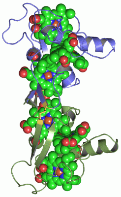
Asymmetric Unit (10, 10)
| No. | Name | Evidence | Residues | Description |
|---|
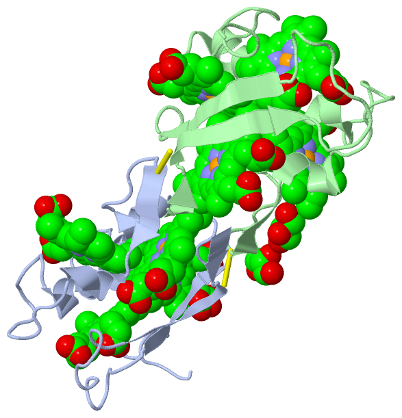
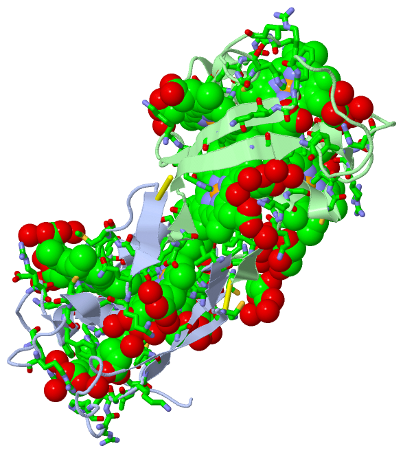
| 01 | AC1 | SOFTWARE | LEU A:1 , MET A:26 , HIS A:28 , HIS A:31 , VAL A:34 , SER A:35 , CYS A:36 , CYS A:39 , HIS A:40 , PHE A:49 , LYS A:51 , CYS A:52 , HEC A:113 , HOH A:2122 , HOH A:2123 , HOH A:2124 , HOH A:2125 , HOH A:2126 , PHE B:43 | BINDING SITE FOR RESIDUE HEC A 111 |
| 02 | AC2 | SOFTWARE | CYS A:39 , HIS A:40 , HIS A:41 , MET A:42 , GLN A:50 , CYS A:52 , CYS A:55 , HIS A:56 , GLU A:65 , ARG A:66 , LYS A:70 , ALA A:71 , SER A:74 , ILE A:78 , SER A:79 , HOH A:2127 , HOH A:2128 , HOH A:2129 , HOH A:2130 , HOH A:2131 , ARG B:20 , LEU B:100 | BINDING SITE FOR RESIDUE HEC A 112 |
| 03 | AC3 | SOFTWARE | ILE A:9 , LYS A:30 , HIS A:31 , CYS A:39 , GLU A:77 , ILE A:78 , SER A:79 , CYS A:80 , CYS A:83 , HIS A:84 , MET A:87 , GLN A:93 , THR A:94 , GLY A:95 , PRO A:96 , HEC A:111 , HOH A:2132 , HOH A:2133 , HOH A:2134 , HOH A:2136 | BINDING SITE FOR RESIDUE HEC A 113 |
| 04 | AC4 | SOFTWARE | ILE A:9 , HIS A:18 , PHE A:21 , GLY A:22 , LYS A:23 , VAL A:24 , ARG A:59 , ARG A:62 , PHE A:68 , TYR A:69 , TRP A:72 , HIS A:73 , CYS A:80 , ARG A:81 , HIS A:84 , PRO A:96 , ILE A:97 , GLY A:98 , CYS A:99 , CYS A:103 , HIS A:104 , HOH A:2137 , HOH A:2138 , HOH A:2139 , GLN B:50 | BINDING SITE FOR RESIDUE HEC A 114 |
| 05 | AC5 | SOFTWARE | LEU A:1 , ASP B:2 , VAL B:3 , ILE B:9 , MET B:26 , HIS B:28 , HIS B:31 , VAL B:34 , SER B:35 , CYS B:36 , CYS B:39 , HIS B:40 , PHE B:49 , LYS B:51 , CYS B:52 , HEC B:113 , HOH B:2127 , HOH B:2128 , HOH B:2129 , HOH B:2130 , HOH B:2131 | BINDING SITE FOR RESIDUE HEC B 111 |
| 06 | AC6 | SOFTWARE | ARG A:20 , LEU A:100 , CYS B:39 , HIS B:40 , HIS B:41 , MET B:42 , CYS B:52 , CYS B:55 , HIS B:56 , GLU B:65 , ARG B:66 , LYS B:70 , ALA B:71 , SER B:74 , ILE B:78 , SER B:79 , GOL B:115 , HOH B:2132 , HOH B:2133 | BINDING SITE FOR RESIDUE HEC B 112 |
| 07 | AC7 | SOFTWARE | LYS B:30 , HIS B:31 , CYS B:39 , ILE B:78 , SER B:79 , CYS B:80 , CYS B:83 , HIS B:84 , MET B:87 , THR B:94 , GLY B:95 , PRO B:96 , HEC B:111 , HOH B:2134 , HOH B:2135 | BINDING SITE FOR RESIDUE HEC B 113 |
| 08 | AC8 | SOFTWARE | GLN A:50 , ILE B:9 , ALA B:11 , PHE B:21 , GLY B:22 , LYS B:23 , VAL B:24 , ARG B:59 , PHE B:68 , TYR B:69 , TRP B:72 , HIS B:73 , CYS B:80 , ARG B:81 , HIS B:84 , PRO B:96 , ILE B:97 , GLY B:98 , CYS B:99 , CYS B:103 , HIS B:104 , HOH B:2025 , HOH B:2136 , HOH B:2137 | BINDING SITE FOR RESIDUE HEC B 114 |
| 09 | AC9 | SOFTWARE | PHE A:21 , ARG A:62 , MET B:42 , GLN B:50 , HEC B:112 , GOL B:116 , HOH B:2064 , HOH B:2067 , HOH B:2069 , HOH B:2138 | BINDING SITE FOR RESIDUE GOL B 115 |
| 10 | BC1 | SOFTWARE | CYS A:5 , HIS A:18 , ARG A:32 , ARG A:62 , ASP B:44 , CYS B:46 , GOL B:115 , HOH B:2139 , HOH B:2140 , HOH B:2141 | BINDING SITE FOR RESIDUE GOL B 116 |
|
 Description