|
|
|
|
 Description
|
|
Compounds
|
||||||||||||||||||||||||||||||||||||||||
Chains, Units
Summary Information (see also Sequences/Alignments below) |
Ligands, Modified Residues, Ions (0, 0)| (no "Ligand,Modified Residues,Ions" information available for 1GHH) |
Sites (0, 0)| (no "Site" information available for 1GHH) |
SS Bonds (0, 0)| (no "SS Bond" information available for 1GHH) |
Cis Peptide Bonds (0, 0)| (no "Cis Peptide Bond" information available for 1GHH) |
SAPs(SNPs)/Variants (0, 0)| (no "SAP(SNP)/Variant" information available for 1GHH) |
PROSITE Motifs (0, 0)| (no "PROSITE Motif" information available for 1GHH) |
Exons (0, 0)| (no "Exon" information available for 1GHH) |
Sequences/Alignments
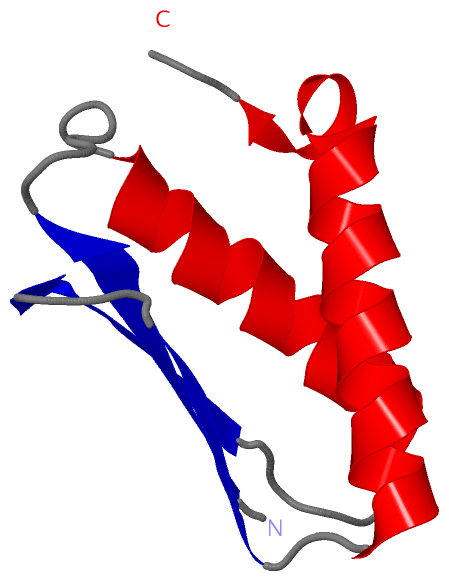
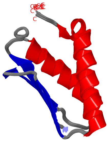
NMR StructureChain A from PDB Type:PROTEIN Length:81 aligned with DINI_ECOLI | P0ABR1 from UniProtKB/Swiss-Prot Length:81 Alignment length:81 10 20 30 40 50 60 70 80 DINI_ECOLI 1 MRIEVTIAKTSPLPAGAIDALAGELSRRIQYAFPDNEGHVSVRYAAANNLSVIGATKEDKQRISEILQETWESADDWFVSE 81 SCOP domains d1ghha_ A: DNA damage-inducible protein DinI SCOP domains CATH domains 1ghhA00 A:1-81 Protein Binding, Dini Protein; Chain A CATH domains Pfam domains --------------------------------------------------------------------------------- Pfam domains SAPs(SNPs) --------------------------------------------------------------------------------- SAPs(SNPs) PROSITE --------------------------------------------------------------------------------- PROSITE Transcript --------------------------------------------------------------------------------- Transcript 1ghh A 1 MRIEVTIAKTSPLPAGAIDALAGELSRRIQYAFPDNEGHVSVRYAAANNLSVIGATKEDKQRISEILQETWESADDWFVSE 81 10 20 30 40 50 60 70 80
|
||||||||||||||||||||
SCOP Domains (1, 1)
NMR Structure
 |
CATH Domains (1, 1)
NMR Structure
|
Pfam Domains (0, 0)| (no "Pfam Domain" information available for 1GHH) |
Gene Ontology (4, 4)|
NMR Structure(hide GO term definitions) Chain A (DINI_ECOLI | P0ABR1)
|
||||||||||||||||||||||||||||||||||||
Interactive Views
|
||||||||||||||||||||||||||||||||||||||||||||||||||||||||||||||||||||||||||||||||||||||||||||||||||||||||||||||||||||
Still Images
|
||||||||||||||||
Databases
|
||||||||||||||||||||||||||||||||||||||||||||||||||||||||||||||||||||||||||||||||||||||||||||||||||||||||||||||||||||||||||||||||||||||||||||||||||||||||||||||||
Analysis Tools
|
|||||||||||||||||||||||||||||||||||||||||||||||||||||||||||||
Entries Sharing at Least One Protein Chain (UniProt ID)
Related Entries Specified in the PDB File
|
|