|
|
|
|
 Description
|
|
Compounds
|
||||||||||||||||||||||||||||||||||||||||
Chains, Units
Summary Information (see also Sequences/Alignments below) |
Ligands, Modified Residues, Ions (2, 2)| Asymmetric Unit (2, 2) Biological Unit 1 (1, 2) Biological Unit 2 (1, 2) |
Sites (2, 2)
Asymmetric Unit (2, 2)
|
SS Bonds (0, 0)| (no "SS Bond" information available for 1G5T) |
Cis Peptide Bonds (0, 0)| (no "Cis Peptide Bond" information available for 1G5T) |
SAPs(SNPs)/Variants (0, 0)| (no "SAP(SNP)/Variant" information available for 1G5T) |
PROSITE Motifs (0, 0)| (no "PROSITE Motif" information available for 1G5T) |
Exons (0, 0)| (no "Exon" information available for 1G5T) |
Sequences/Alignments
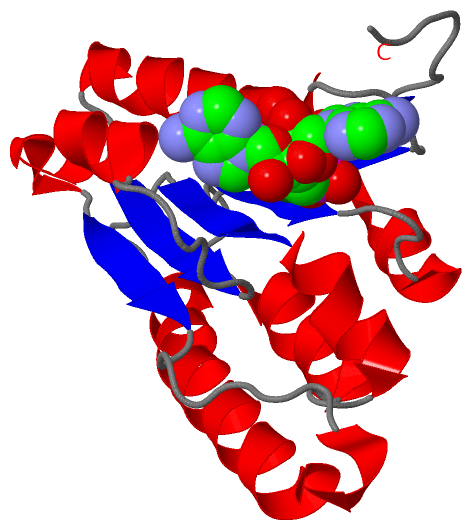
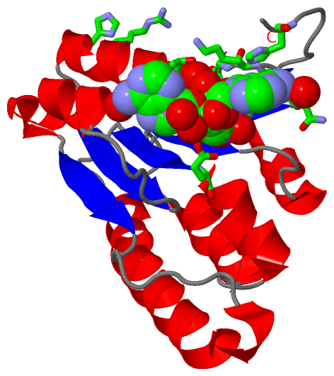
Asymmetric UnitChain A from PDB Type:PROTEIN Length:157 aligned with BTUR_SALTY | P31570 from UniProtKB/Swiss-Prot Length:196 Alignment length:157 36 46 56 66 76 86 96 106 116 126 136 146 156 166 176 BTUR_SALTY 27 ERGIIIVFTGNGKGKTTAAFGTAARAVGHGKNVGVVQFIKGTWPNGERNLLEPHGVEFQVMATGFTWETQNREADTAACMAVWQHGKRMLADPLLDMVVLDELTYMVAYDYLPLEEVISALNARPGHQTVIITGRGCHRDILDLADTVSELRPVKHA 183 SCOP domains d1g5ta_ A: ATP:corrinoid adenosyltransferase CobA SCOP domains CATH domains 1g5tA00 A:27-183 P-loop containing nucleotide triphosphate hydrolases CATH domains Pfam domains ------------------------------------------------------------------------------------------------------------------------------------------------------------- Pfam domains SAPs(SNPs) ------------------------------------------------------------------------------------------------------------------------------------------------------------- SAPs(SNPs) PROSITE ------------------------------------------------------------------------------------------------------------------------------------------------------------- PROSITE Transcript ------------------------------------------------------------------------------------------------------------------------------------------------------------- Transcript 1g5t A 27 ERGIIIVFTGNGKGKTTAAFGTAARAVGHGKNVGVVQFIKGTWPNGERNLLEPHGVEFQVMATGFTWETQNREADTAACMAVWQHGKRMLADPLLDMVVLDELTYMVAYDYLPLEEVISALNARPGHQTVIITGRGCHRDILDLADTVSELRPVKHA 183 36 46 56 66 76 86 96 106 116 126 136 146 156 166 176
|
||||||||||||||||||||
SCOP Domains (1, 1)
Asymmetric Unit
 |
CATH Domains (1, 1)
Asymmetric Unit
|
Pfam Domains (0, 0)| (no "Pfam Domain" information available for 1G5T) |
Gene Ontology (7, 7)|
Asymmetric Unit(hide GO term definitions) Chain A (BTUR_SALTY | P31570)
|
||||||||||||||||||||||||||||||||||||||||||||||||||||||||||||
Interactive Views
|
|||||||||||||||||||||||||||||||||||||||||||||||||||||||||||||||||||||||||||||||||||||||||||||||||||||||||||||||||||||||||||||||||||||||||||||||||||||||||||
Still Images
|
||||||||||||||||
Databases
|
||||||||||||||||||||||||||||||||||||||||||||||||||||||||||||||||||||||||||||||||||||||||||||||||||||||||||||||||||||||||||||||||||||||||||||||||||||||||||||||||
Analysis Tools
|
|||||||||||||||||||||||||||||||||||||||||||||||||||||||||||||
Entries Sharing at Least One Protein Chain (UniProt ID)
Related Entries Specified in the PDB File
|
|