|
|
|
|
 Description
|
|
Compounds
|
||||||||||||||||||||||||||||||||
Chains, Units
Summary Information (see also Sequences/Alignments below) |
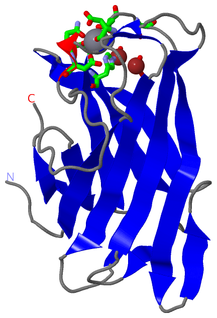
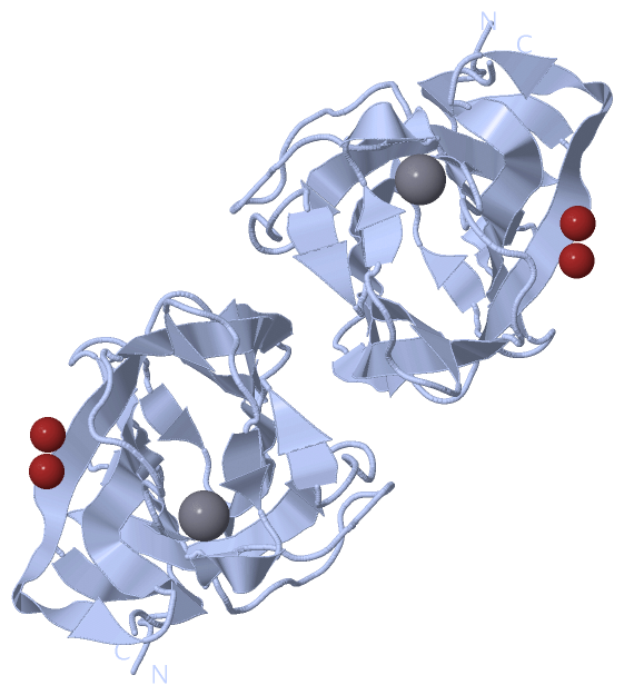
Ligands, Modified Residues, Ions (2, 3)| Asymmetric Unit (2, 3) Biological Unit 1 (0, 0) Biological Unit 2 (0, 0) |
Sites (3, 3)
Asymmetric Unit (3, 3)
|
SS Bonds (0, 0)| (no "SS Bond" information available for 1G43) |
Cis Peptide Bonds (1, 1)
Asymmetric Unit
|
||||||||
SAPs(SNPs)/Variants (0, 0)| (no "SAP(SNP)/Variant" information available for 1G43) |
PROSITE Motifs (0, 0)| (no "PROSITE Motif" information available for 1G43) |
Exons (0, 0)| (no "Exon" information available for 1G43) |
Sequences/Alignments
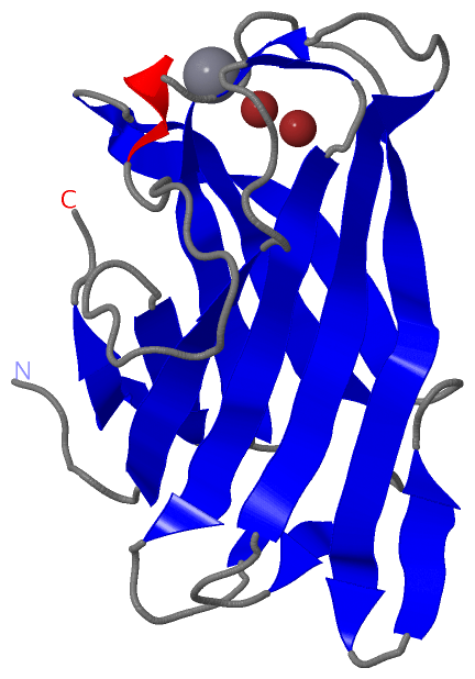
Asymmetric UnitChain A from PDB Type:PROTEIN Length:160 aligned with Q45996_9FIRM | Q45996 from UniProtKB/TrEMBL Length:1546 Alignment length:160 37 47 57 67 77 87 97 107 117 127 137 147 157 167 177 187 Q45996_9FIRM 28 AGTGVVSVQFNNGSSPASSNSIYARFKVTNTSGSPINLADLKLRYYYTQDADKPLTFWCDHAGYMSGSNYIDATSKVTGSFKAVSPAVTNADHYLEVALNSDAGSLPAGGSIEIQTRFARNDWSNFDQSNDWSYTAAGSYMDWQKISAFVGGTLAYGSTP 187 SCOP domains d1g43a_ A: Cellusomal scaffolding protein A, scaffoldin SCOP domains CATH domains 1g43A00 A:1-160 [code=2.60.40.710, no name defined] CATH domains Pfam domains ---------------------------------------------------------------------------------------------------------------------------------------------------------------- Pfam domains SAPs(SNPs) ---------------------------------------------------------------------------------------------------------------------------------------------------------------- SAPs(SNPs) PROSITE ---------------------------------------------------------------------------------------------------------------------------------------------------------------- PROSITE Transcript ---------------------------------------------------------------------------------------------------------------------------------------------------------------- Transcript 1g43 A 1 AGTGVVSVQFNNGSSPASSNSIYARFKVTNTSGSPINLADLKLRYYYTQDADKPLTFWCDHAGYMSGSNYIDATSKVTGSFKAVSPAVTNADHYLEVALNSDAGSLPAGGSIEIQTRFARNDWSNFDQSNDWSYTAAGSYMDWQKISAFVGGTLAYGSTP 160 10 20 30 40 50 60 70 80 90 100 110 120 130 140 150 160
|
||||||||||||||||||||
SCOP Domains (1, 1)
Asymmetric Unit
 |
CATH Domains (1, 1)
Asymmetric Unit
|
Pfam Domains (0, 0)| (no "Pfam Domain" information available for 1G43) |
Gene Ontology (5, 5)|
Asymmetric Unit(hide GO term definitions) Chain A (Q45996_9FIRM | Q45996)
|
||||||||||||||||||||||||||||||||||||||||||
Interactive Views
|
|||||||||||||||||||||||||||||||||||||||||||||||||||||||||||||||||||||||||||||||||||||||||||||||||||||||||||||||||||||||||||||||||||||||||||||||||||||||||||||||||||
Still Images
|
||||||||||||||||
Databases
|
||||||||||||||||||||||||||||||||||||||||||||||||||||||||||||||||||||||||||||||||||||||||||||||||||||||||||||||||||||||||||||||||||||||||||||||||||||||||||||||||
Analysis Tools
|
|||||||||||||||||||||||||||||||||||||||||||||||||||||||||||||
Entries Sharing at Least One Protein Chain (UniProt ID)
Related Entries Specified in the PDB File
|
|