| molecular function |
|---|
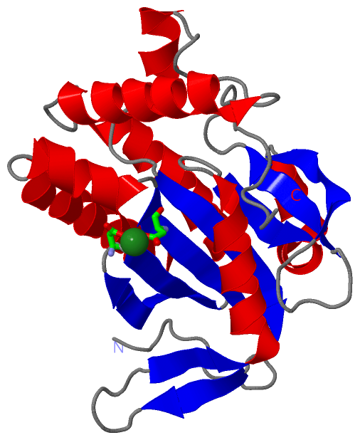
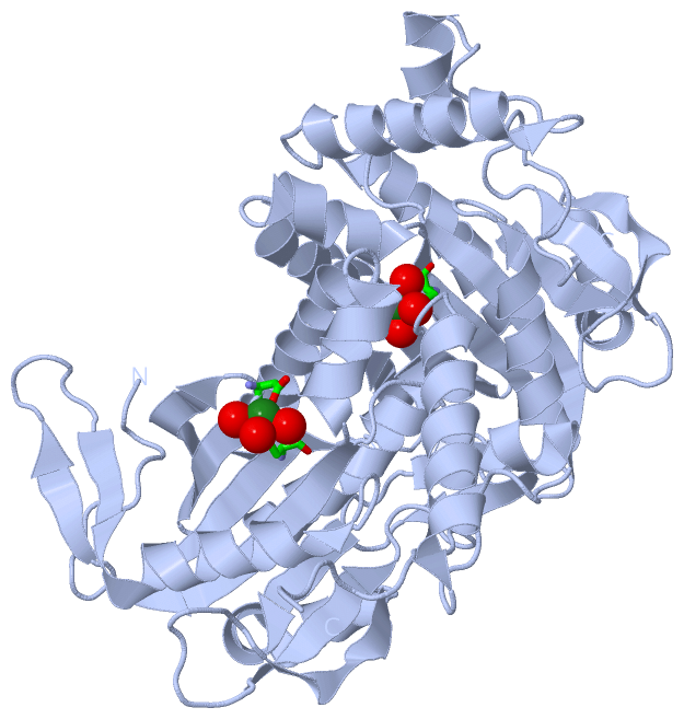
| | GO:0005524 | | ATP binding | | Interacting selectively and non-covalently with ATP, adenosine 5'-triphosphate, a universally important coenzyme and enzyme regulator. |
| | GO:0004016 | | adenylate cyclase activity | | Catalysis of the reaction: ATP = 3',5'-cyclic AMP + diphosphate. |
| | GO:0016829 | | lyase activity | | Catalysis of the cleavage of C-C, C-O, C-N and other bonds by other means than by hydrolysis or oxidation, or conversely adding a group to a double bond. They differ from other enzymes in that two substrates are involved in one reaction direction, but only one in the other direction. When acting on the single substrate, a molecule is eliminated and this generates either a new double bond or a new ring. |
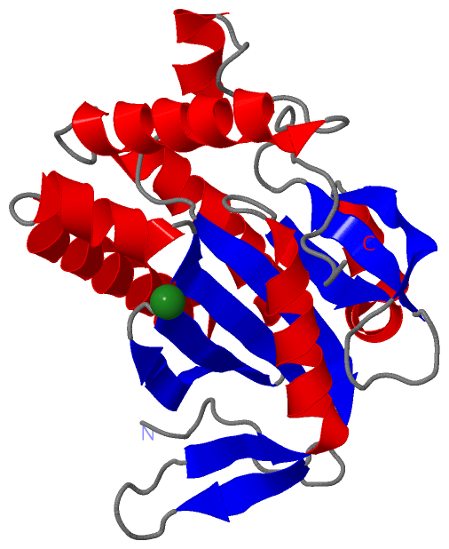
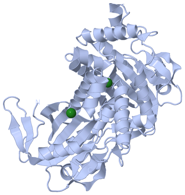
| | GO:0046872 | | metal ion binding | | Interacting selectively and non-covalently with any metal ion. |
| | GO:0000166 | | nucleotide binding | | Interacting selectively and non-covalently with a nucleotide, any compound consisting of a nucleoside that is esterified with (ortho)phosphate or an oligophosphate at any hydroxyl group on the ribose or deoxyribose. |
| | GO:0016849 | | phosphorus-oxygen lyase activity | | Catalysis of the cleavage of a phosphorus-oxygen bond by other means than by hydrolysis or oxidation, or conversely adding a group to a double bond. |
| biological process |
|---|
| | GO:0006171 | | cAMP biosynthetic process | | The chemical reactions and pathways resulting in the formation of the nucleotide cAMP (cyclic AMP, adenosine 3',5'-cyclophosphate). |
| | GO:0009190 | | cyclic nucleotide biosynthetic process | | The chemical reactions and pathways resulting in the formation of a cyclic nucleotide, a nucleotide in which the phosphate group is in diester linkage to two positions on the sugar residue. |
| | GO:0035556 | | intracellular signal transduction | | The process in which a signal is passed on to downstream components within the cell, which become activated themselves to further propagate the signal and finally trigger a change in the function or state of the cell. |
| cellular component |
|---|
| | GO:0016021 | | integral component of membrane | | The component of a membrane consisting of the gene products and protein complexes having at least some part of their peptide sequence embedded in the hydrophobic region of the membrane. |
| | GO:0005622 | | intracellular | | The living contents of a cell; the matter contained within (but not including) the plasma membrane, usually taken to exclude large vacuoles and masses of secretory or ingested material. In eukaryotes it includes the nucleus and cytoplasm. |
| | GO:0016020 | | membrane | | A lipid bilayer along with all the proteins and protein complexes embedded in it an attached to it. |
 Description