|
|
|
|
 Description
|
|
Compounds
|
||||||||||||||||||||||||||||||||||||||||||||
Chains, Units
Summary Information (see also Sequences/Alignments below) |
Ligands, Modified Residues, Ions (0, 0)| (no "Ligand,Modified Residues,Ions" information available for 1FOV) |
Sites (0, 0)| (no "Site" information available for 1FOV) |
SS Bonds (1, 1)
NMR Structure
|
||||||||
Cis Peptide Bonds (1, 20)
NMR Structure
|
||||||||||
SAPs(SNPs)/Variants (0, 0)| (no "SAP(SNP)/Variant" information available for 1FOV) |
PROSITE Motifs (1, 1)
NMR Structure (1, 1)
|
||||||||||||||||||||||||
Exons (0, 0)| (no "Exon" information available for 1FOV) |
Sequences/Alignments
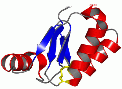
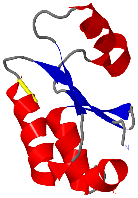
NMR StructureChain A from PDB Type:PROTEIN Length:82 aligned with GLRX3_ECOLI | P0AC62 from UniProtKB/Swiss-Prot Length:83 Alignment length:82 11 21 31 41 51 61 71 81 GLRX3_ECOLI 2 ANVEIYTKETCPYCHRAKALLSSKGVSFQELPIDGNAAKREEMIKRSGRTTVPQIFIDAQHIGGCDDLYALDARGGLDPLLK 83 SCOP domains d1fova_ A: Glutaredoxin (Grx, thioltransferase) SCOP domains CATH domains 1fovA00 A:1-82 Glutaredoxin CATH domains Pfam domains ---------------------------------------------------------------------------------- Pfam domains SAPs(SNPs) ---------------------------------------------------------------------------------- SAPs(SNPs) PROSITE ----GLUTAREDOXIN_1 ------------------------------------------------------------- PROSITE Transcript ---------------------------------------------------------------------------------- Transcript 1fov A 1 ANVEIYTKETCPYCHRAKALLSSKGVSFQELPIDGNAAKREEMIKRSGRTTVPQIFIDAQHIGGYDDLYALDARGGLDPLLK 82 10 20 30 40 50 60 70 80
|
||||||||||||||||||||
SCOP Domains (1, 1)
NMR Structure
 |
CATH Domains (1, 1)
NMR Structure
|
Pfam Domains (0, 0)| (no "Pfam Domain" information available for 1FOV) |
Gene Ontology (7, 7)|
NMR Structure(hide GO term definitions) Chain A (GLRX3_ECOLI | P0AC62)
|
||||||||||||||||||||||||||||||||||||||||||||||||||||||||||||
Interactive Views
|
|||||||||||||||||||||||||||||||||||||||||||||||||||||||||||||||||||||||||||||||||||||||||||||||||||||||||||||||||||||
Still Images
|
||||||||||||||||
Databases
|
||||||||||||||||||||||||||||||||||||||||||||||||||||||||||||||||||||||||||||||||||||||||||||||||||||||||||||||||||||||||||||||||||||||||||||||||||||||||||||||||
Analysis Tools
|
|||||||||||||||||||||||||||||||||||||||||||||||||||||||||||||
Entries Sharing at Least One Protein Chain (UniProt ID)
Related Entries Specified in the PDB File
|
|