|
|
|
|
 Description
|
|
Compounds
|
||||||||||||||||||||||||||||||||||||||||||||||||||
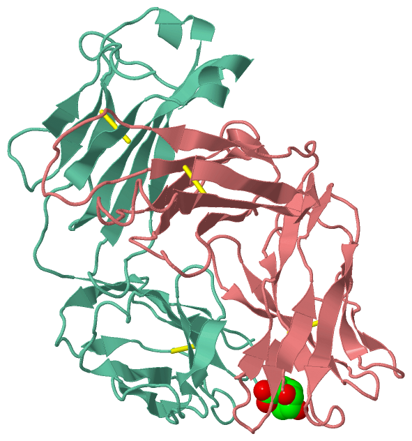
Chains, Units
Summary Information (see also Sequences/Alignments below) |
Ligands, Modified Residues, Ions (1, 1)
Asymmetric/Biological Unit (1, 1)
|
Sites (1, 1)
Asymmetric Unit (1, 1)
|
SS Bonds (4, 4)
Asymmetric/Biological Unit
|
||||||||||||||||||||
Cis Peptide Bonds (7, 7)
Asymmetric/Biological Unit
|
||||||||||||||||||||||||||||||||
SAPs(SNPs)/Variants (0, 0)| (no "SAP(SNP)/Variant" information available for 1FIG) |
PROSITE Motifs (0, 0)| (no "PROSITE Motif" information available for 1FIG) |
Exons (0, 0)| (no "Exon" information available for 1FIG) |
Sequences/Alignments
Asymmetric/Biological Unit
Chain H from PDB Type:PROTEIN Length:216
SCOP domains d1figh1 H:1-113 Immunoglobulin heavy chain variable domain, VH d1figh2 H:114-223 Immunoglobulin heavy chain gamma constant domain 1, CH1-gamma SCOP domains
CATH domains 1figH01 H:1-114 Immunoglobulins 1figH02 H:115-223 Immunoglobulins CATH domains
Pfam domains ------------------------------------------------------------------------------------------------------------------------------------------------------------------------------------------------------------------------ Pfam domains
SAPs(SNPs) ------------------------------------------------------------------------------------------------------------------------------------------------------------------------------------------------------------------------ SAPs(SNPs)
PROSITE ------------------------------------------------------------------------------------------------------------------------------------------------------------------------------------------------------------------------ PROSITE
Transcript ------------------------------------------------------------------------------------------------------------------------------------------------------------------------------------------------------------------------ Transcript
1fig H 1 DVQLQQSGPELEKPGASVKISCKASGFSLPGHNINWIVQRNGKSLEWIGNIDPYYGGTNFNPKFKGKATLTVDKSSSTLYMHLTSLQSEDSAVYYCARRRDGNYGFTYWGQGTLVTVSAAKTTPPSVYPLAPGSAAQTNSMVTLGCLVKGYFPEPVTVTWNSGSLSSGVHTFPAVLQSDLYTLSSSVTVPSSTWPSETVTCNVAHPASSTKVDKKI 223
10 20 30 40 50 | 59 69 79 ||| 86 96 || 104 114 124 ||136 146 157| 172 184 194 || || 206| 217
52A 82A|| 100A| 130| 154||| 169| 180| 196| || 206|
82B| 100B 133 156|| 171 183 198 || 208
82C 157| 200|
162 202
Chain L from PDB Type:PROTEIN Length:215
SCOP domains d1figl1 L:1-108 Immunoglobulin light chain kappa variable domain, VL-kappa d1figl2 L:109-214 Immunoglobulin light chain kappa constant domain, CL-kappa SCOP domains
CATH domains 1figL01 L:1-108 Immunoglobulins 1figL02 L:109-211 Immunoglobulins --- CATH domains
Pfam domains ----------------------------------------------------------------------------------------------------------------------------------------------------------------------------------------------------------------------- Pfam domains
SAPs(SNPs) ----------------------------------------------------------------------------------------------------------------------------------------------------------------------------------------------------------------------- SAPs(SNPs)
PROSITE ----------------------------------------------------------------------------------------------------------------------------------------------------------------------------------------------------------------------- PROSITE
Transcript ----------------------------------------------------------------------------------------------------------------------------------------------------------------------------------------------------------------------- Transcript
1fig L 1 ENVLTQSPAIMSASPGEKVTMACRASSSVSSTYLHWYQQKSGASPKLLIYSTSNLASGVPARFSGSGSGTSYSLTISSVEAEDAATYYCQQYSGYPLTFGAGTKLELKRADAAPTVSIFPPSSEQLTSGGASVVCFLNNFYPKDINVKWKIDGSERQNGVLNSWTDQDSKDSTYSMSSTLTLTKDEYERHNSYTCEATHKTSTSPIVKSFNRNEC 214
10 20 |29 39 49 59 69 79 89 99 109 119 129 139 149 159 169 179 189 199 209
27A
|
||||||||||||||||||||
SCOP Domains (4, 4)
Asymmetric/Biological Unit
 |
CATH Domains (1, 4)
Asymmetric/Biological Unit
|
Pfam Domains (0, 0)| (no "Pfam Domain" information available for 1FIG) |
Gene Ontology (0, 0)|
Asymmetric/Biological Unit(hide GO term definitions)
(no "Gene Ontology" information available for 1FIG)
|
Interactive Views
|
|||||||||||||||||||||||||||||||||||||||||||||||||||||||||||||||||||||||||||||||||||||||||||||||||||||||||||||||||||||||||||||||||||||||||||||||||||||||||||||||||
Still Images
|
||||||||||||||||
Databases
|
||||||||||||||||||||||||||||||||||||||||||||||||||||||||||||||||||||||||||||||||||||||||||||||||||||||||||||||||||||||||||||||||||||||||||||||||||||||||||||||||
Analysis Tools
|
|||||||||||||||||||||||||||||||||||||||||||||||||||||||||||||
Entries Sharing at Least One Protein Chain (UniProt ID)
Related Entries Specified in the PDB File
|
|