| molecular function |
|---|
| | GO:0005524 | | ATP binding | | Interacting selectively and non-covalently with ATP, adenosine 5'-triphosphate, a universally important coenzyme and enzyme regulator. |
| | GO:0016887 | | ATPase activity | | Catalysis of the reaction: ATP + H2O = ADP + phosphate + 2 H+. May or may not be coupled to another reaction. |
| | GO:0003677 | | DNA binding | | Any molecular function by which a gene product interacts selectively and non-covalently with DNA (deoxyribonucleic acid). |
| | GO:0016787 | | hydrolase activity | | Catalysis of the hydrolysis of various bonds, e.g. C-O, C-N, C-C, phosphoric anhydride bonds, etc. Hydrolase is the systematic name for any enzyme of EC class 3. |
| | GO:0046872 | | metal ion binding | | Interacting selectively and non-covalently with any metal ion. |
| | GO:0000166 | | nucleotide binding | | Interacting selectively and non-covalently with a nucleotide, any compound consisting of a nucleoside that is esterified with (ortho)phosphate or an oligophosphate at any hydroxyl group on the ribose or deoxyribose. |
| | GO:0005515 | | protein binding | | Interacting selectively and non-covalently with any protein or protein complex (a complex of two or more proteins that may include other nonprotein molecules). |
| biological process |
|---|
| | GO:0015074 | | DNA integration | | The process in which a segment of DNA is incorporated into another, usually larger, DNA molecule such as a chromosome. |
| | GO:0006260 | | DNA replication | | The cellular metabolic process in which a cell duplicates one or more molecules of DNA. DNA replication begins when specific sequences, known as origins of replication, are recognized and bound by initiation proteins, and ends when the original DNA molecule has been completely duplicated and the copies topologically separated. The unit of replication usually corresponds to the genome of the cell, an organelle, or a virus. The template for replication can either be an existing DNA molecule or RNA. |
| | GO:0032196 | | transposition | | Any process involved in mediating the movement of discrete segments of DNA between nonhomologous sites. |
| | GO:0006313 | | transposition, DNA-mediated | | Any process involved in a type of transpositional recombination which occurs via a DNA intermediate. |
| | GO:0039693 | | viral DNA genome replication | | The replication of a viral DNA genome. |
| cellular component |
|---|
| | GO:0030430 | | host cell cytoplasm | | The cytoplasm of a host cell. |
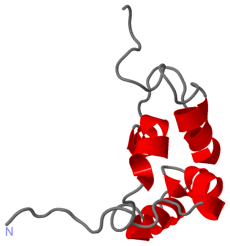
 Description
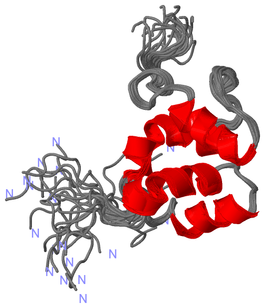