Show PDB file:   
         Plain Text   HTML   (compressed file size)
QuickSearch:   
by PDB,NDB,UniProt,PROSITE Code or Search Term(s)  
(-)Asymmetric Unit
(-)Biological Unit 1
(-)Biological Unit 2
collapse expand < >
Image Asymmetric Unit
Asymmetric Unit  (Jmol Viewer)
Image Biological Unit 1
Biological Unit 1  (Jmol Viewer)
Image Biological Unit 2
Biological Unit 2  (Jmol Viewer)

(-) Description

Title :  STRUCTURE OF THE SINGLE-DOMAIN CAMELID ANTIBODY CAB-CA05
 
Authors :  K. Decanniere, S. Muyldermans, L. Wyns
Date :  30 May 00  (Deposition) - 08 Jun 00  (Release) - 24 Feb 09  (Revision)
Method :  X-RAY DIFFRACTION
Resolution :  2.10
Chains :  Asym. Unit :  K,L
Biol. Unit 1:  K  (1x)
Biol. Unit 2:  L  (1x)
Keywords :  Immunoglobulin Fold, Immune System (Keyword Search: [Gene Ontology, PubMed, Web (Google))
 
Reference :  K. Decanniere, S. Muyldermans, L. Wyns
Canonical Antigen-Binding Loop Structures In Immunoglobulins: More Structures, More Canonical Classes?
J. Mol. Biol. V. 300 83 2000
PubMed-ID: 10864500  |  Reference-DOI: 10.1006/JMBI.2000.3839
(for further references see the PDB file header)

(-) Compounds

Molecule 1 - ANTIBODY HEAVY CHAIN
    CellLYMPHOCYTES
    ChainsK, L
    EngineeredYES
    Expression SystemESCHERICHIA COLI
    Expression System Taxid562
    Organism CommonARABIAN CAMEL
    Organism ScientificCAMELUS DROMEDARIUS
    Organism Taxid9838
    SynonymVHH

 Structural Features

(-) Chains, Units

  12
Asymmetric Unit KL
Biological Unit 1 (1x)K 
Biological Unit 2 (1x) L

Summary Information (see also Sequences/Alignments below)

(-) Ligands, Modified Residues, Ions  (0, 0)

(no "Ligand,Modified Residues,Ions" information available for 1F2X)

(-) Sites  (0, 0)

(no "Site" information available for 1F2X)

(-) SS Bonds  (4, 4)

Asymmetric Unit
No.Residues
1K:822 -K:894
2K:833 -K:905
3L:1022 -L:1094
4L:1033 -L:1105

(-) Cis Peptide Bonds  (0, 0)

(no "Cis Peptide Bond" information available for 1F2X)

 Sequence-Structure Mapping

(-) SAPs(SNPs)/Variants  (0, 0)

(no "SAP(SNP)/Variant" information available for 1F2X)

(-) PROSITE Motifs  (0, 0)

(no "PROSITE Motif" information available for 1F2X)

(-) Exons   (0, 0)

(no "Exon" information available for 1F2X)

(-) Sequences/Alignments

Asymmetric Unit
   Reformat: Number of residues per line =  ('0' or empty: single-line sequence representation)
  Number of residues per labelling interval =   
  UniProt sequence: complete  aligned part    
   Show mapping: SCOP domains CATH domains Pfam domains Secondary structure (by author)
SAPs(SNPs) PROSITE motifs Exons
(details for a mapped element are shown in a popup box when the mouse pointer rests over it)
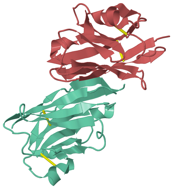
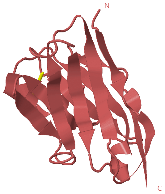
Chain K from PDB  Type:PROTEIN  Length:126
                                                                                                                                                               
               SCOP domains d1f2xk_ K: Camelid IG heavy chain variable domain, VHh                                                                         SCOP domains
               CATH domains 1f2xK00 K:801-926 Immunoglobulins                                                                                              CATH domains
               Pfam domains ------------------------------------------------------------------------------------------------------------------------------ Pfam domains
         Sec.struct. author ..eeeee..eeee....eeeeeeee........eeeeee......eeeeeee..eeee.......eeeeee....eeeeee...hhhhheeeeeee....hhhhhhhhhhhhh.ee...eeeeee. Sec.struct. author
                 SAPs(SNPs) ------------------------------------------------------------------------------------------------------------------------------ SAPs(SNPs)
                    PROSITE ------------------------------------------------------------------------------------------------------------------------------ PROSITE
                 Transcript ------------------------------------------------------------------------------------------------------------------------------ Transcript
                1f2x K  801 QVQLVESGGGSVQAGGSLRLSCAASGYTVSTYCMGWFRQAPGKEREGVATILGGSTYYGDSVKGRFTISQDNAKNTVYLQMNSLKPEDTAIYYCAGSTVASTGWCSRLRPYDYHYRGQGTQVTVSS  926
                                   810       820       830       840       850       860       870       880       890       900       910       920      

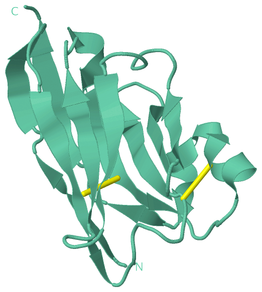
Chain L from PDB  Type:PROTEIN  Length:126
                                                                                                                                                               
               SCOP domains d1f2xl_ L: Camelid IG heavy chain variable domain, VHh                                                                         SCOP domains
               CATH domains 1f2xL00 L:1002-1127 Immunoglobulins                                                                                            CATH domains
               Pfam domains ------------------------------------------------------------------------------------------------------------------------------ Pfam domains
         Sec.struct. author .eeeee..eeee....eeeeeeee........eeeeee......eeeeeee..eeee.......eeeeee....eeeeee...hhhhheeeeeee....hhhhhhhhhhhhh.ee...eeeeee.. Sec.struct. author
                 SAPs(SNPs) ------------------------------------------------------------------------------------------------------------------------------ SAPs(SNPs)
                    PROSITE ------------------------------------------------------------------------------------------------------------------------------ PROSITE
                 Transcript ------------------------------------------------------------------------------------------------------------------------------ Transcript
                1f2x L 1002 VQLVESGGGSVQAGGSLRLSCAASGYTVSTYCMGWFRQAPGKEREGVATILGGSTYYGDSVKGRFTISQDNAKNTVYLQMNSLKPEDTAIYYCAGSTVASTGWCSRLRPYDYHYRGQGTQVTVSSR 1127
                                  1011      1021      1031      1041      1051      1061      1071      1081      1091      1101      1111      1121      

   Legend:   → Mismatch (orange background)
  - → Gap (green background, '-', border residues have a numbering label)
    → Modified Residue (blue background, lower-case, 'x' indicates undefined single-letter code, labelled with number + name)
  x → Chemical Group (purple background, 'x', labelled with number + name, e.g. ACE or NH2)
  extra numbering lines below/above indicate numbering irregularities and modified residue names etc., number ends below/above '|'

 Classification and Annotation

(-) SCOP Domains  (1, 2)

Asymmetric Unit

(-) CATH Domains  (1, 2)

Asymmetric Unit
(-)
Class: Mainly Beta (13760)

(-) Pfam Domains  (0, 0)

(no "Pfam Domain" information available for 1F2X)

(-) Gene Ontology  (0, 0)

Asymmetric Unit(hide GO term definitions)
    (no "Gene Ontology" information available for 1F2X)

 Visualization

(-) Interactive Views

Asymmetric Unit
  Complete Structure
    Jena3D(integrated viewing of ligand, site, SAP, PROSITE, SCOP information)
    WebMol | AstexViewer[tm]@PDBe
(Java Applets, require no local installation except for Java; loading may be slow)
    STRAP
(Java WebStart application, automatic local installation, requires Java; full application with system access!)
    RasMol
(require local installation)
    Molscript (VRML)
(requires installation of a VRML viewer; select preferred view via VRML and generate a mono or stereo PDF format file)
 
  Ligands, Modified Residues, Ions
(no "Ligands, Modified Residues, Ions" information available for 1f2x)
 
  Sites
(no "Sites" information available for 1f2x)
 
  Cis Peptide Bonds
(no "Cis Peptide Bonds" information available for 1f2x)
 
Biological Units
  Complete Structure
    Biological Unit 1  [ Jena3D ]
    Biological Unit 2  [ Jena3D ]

(-) Still Images

Jmol
  protein: cartoon or spacefill or dots and stick; nucleic acid: cartoon and stick; ligands: spacefill; active site: stick
Molscript
  protein, nucleic acid: cartoon; ligands: spacefill; active site: ball and stick

 Databases and Analysis Tools

(-) Databases

Access by PDB/NDB ID
  1f2x
    Family and Domain InformationProDom | SYSTERS
    General Structural InformationGlycoscienceDB | MMDB | NDB | OCA | PDB | PDBe | PDBj | PDBsum | PDBWiki | PQS | PROTEOPEDIA
    Orientation in MembranesOPM
    Protein SurfaceSURFACE
    Secondary StructureDSSP (structure derived) | HSSP (homology derived)
    Structural GenomicsGeneCensus
    Structural NeighboursCE | VAST
    Structure ClassificationCATH | Dali | SCOP
    Validation and Original DataBMRB Data View | BMRB Restraints Grid | EDS | PROCHECK | RECOORD | WHAT_CHECK
 
Access by UniProt ID/Accession number
  (no 'UniProt ID/Accession number' available) |
    Comparative Protein Structure ModelsModBase
    Genomic InformationEnsembl
    Protein-protein InteractionDIP
    Sequence, Family and Domain InformationInterPro | Pfam | SMART | UniProt
 
Access by Enzyme Classificator   (EC Number)
  (no 'Enzyme Classificator' available)
    General Enzyme InformationBRENDA | EC-PDB | Enzyme | IntEnz
    PathwayKEGG | MetaCyc
 
Access by Disease Identifier   (MIM ID)
  (no 'MIM ID' available)
    Disease InformationOMIM
 
Access by GenAge ID
  (no 'GenAge ID' available)
    Age Related InformationGenAge

(-) Analysis Tools

Access by PDB/NDB ID
    Domain InformationXDom
    Interatomic Contacts of Structural UnitsCSU
    Ligand-protein ContactsLPC
    Protein CavitiescastP
    Sequence and Secondary StructurePDBCartoon
    Structure AlignmentSTRAP(Java WebStart application, automatic local installation, requires Java; full application with system access!)
    Structure and Sequence BrowserSTING
 
Access by UniProt ID/Accession number
  (no 'UniProt ID/Accession number' available) |
    Protein Disorder PredictionDisEMBL | FoldIndex | GLOBPLOT

 Related Entries

(-) Entries Sharing at Least One Protein Chain (UniProt ID)

(no "Entries Sharing at Least One Protein Chain" available for 1F2X)

(-) Related Entries Specified in the PDB File

1bzq 1BZQ CONTAINS A SINGLE-DOMAIN ANTIBODY FRAGMENT IN COMPLEX WITH RNASE A.
1hcv 1HCV CONTAINS THE CRYSTAL STRUCTURE OF A LLAMA HEAVY CHAIN VARIABLE DOMAIN.
1mel 1MEL CONTAINS A CAMEL SINGLE-DOMAIN VH ANTIBODY FRAGMENT IN COMPLEX WITH LYSOZYME.