| molecular function |
|---|
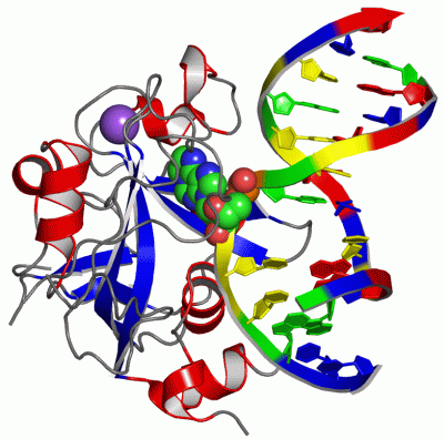
| | GO:0019104 | | DNA N-glycosylase activity | | Catalysis of the removal of damaged bases by cleaving the N-C1' glycosidic bond between the target damaged DNA base and the deoxyribose sugar. The reaction releases a free base and leaves an apurinic/apyrimidinic (AP) site. |
| | GO:0003677 | | DNA binding | | Any molecular function by which a gene product interacts selectively and non-covalently with DNA (deoxyribonucleic acid). |
| | GO:0008725 | | DNA-3-methyladenine glycosylase activity | | Catalysis of the reaction: DNA containing 3-methyladenine + H2O = DNA with abasic site + 3-methyladenine. This reaction is the hydrolysis of DNA by cleavage of the N-C1' glycosidic bond between the damaged DNA 3-methyladenine and the deoxyribose sugar to remove the 3-methyladenine, leaving an abasic site. |
| | GO:0052822 | | DNA-3-methylguanine glycosylase activity | | Catalysis of the reaction: DNA containing 3-methylguanine + H2O = DNA with abasic site + 3-methylguanine. This reaction is the hydrolysis of DNA by cleavage of the N-C1' glycosidic bond between the damaged DNA 3-methylguanine and the deoxyribose sugar to remove the 3-methylguanine, leaving an abasic site. |
| | GO:0052821 | | DNA-7-methyladenine glycosylase activity | | Catalysis of the reaction: DNA containing 7-methyladenine + H2O = DNA with abasic site + 7-methyladenine. This reaction is the hydrolysis of DNA by cleavage of the N-C1' glycosidic bond between the damaged DNA 7-methyladenine and the deoxyribose sugar to remove the 7-methyladenine, leaving an abasic site. |
| | GO:0043916 | | DNA-7-methylguanine glycosylase activity | | Catalysis of the reaction: DNA containing 7-methylguanine + H2O = DNA with abasic site + 7-methylguanine. This reaction is the hydrolysis of DNA by cleavage of the N-C1' glycosidic bond between the damaged DNA 7-methylguanine and the deoxyribose sugar to remove the 7-methylguanine, leaving an abasic site. |
| | GO:0003905 | | alkylbase DNA N-glycosylase activity | | Catalysis of the reaction: DNA with alkylated base + H2O = DNA with abasic site + alkylated base. This reaction is the hydrolysis of DNA by cleavage of the N-C1' glycosidic bond between the target damaged DNA base and the deoxyribose sugar to remove an alkylated base, leaving an apyrimidinic or apurinic site. |
| | GO:0003824 | | catalytic activity | | Catalysis of a biochemical reaction at physiological temperatures. In biologically catalyzed reactions, the reactants are known as substrates, and the catalysts are naturally occurring macromolecular substances known as enzymes. Enzymes possess specific binding sites for substrates, and are usually composed wholly or largely of protein, but RNA that has catalytic activity (ribozyme) is often also regarded as enzymatic. |
| | GO:0003684 | | damaged DNA binding | | Interacting selectively and non-covalently with damaged DNA. |
| | GO:0016787 | | hydrolase activity | | Catalysis of the hydrolysis of various bonds, e.g. C-O, C-N, C-C, phosphoric anhydride bonds, etc. Hydrolase is the systematic name for any enzyme of EC class 3. |
| | GO:0005515 | | protein binding | | Interacting selectively and non-covalently with any protein or protein complex (a complex of two or more proteins that may include other nonprotein molecules). |
| biological process |
|---|
| | GO:0006307 | | DNA dealkylation involved in DNA repair | | The repair of alkylation damage, e.g. the removal of the alkyl group at the O6-position of guanine by O6-alkylguanine-DNA alkyltransferase (AGT). |
| | GO:0006281 | | DNA repair | | The process of restoring DNA after damage. Genomes are subject to damage by chemical and physical agents in the environment (e.g. UV and ionizing radiations, chemical mutagens, fungal and bacterial toxins, etc.) and by free radicals or alkylating agents endogenously generated in metabolism. DNA is also damaged because of errors during its replication. A variety of different DNA repair pathways have been reported that include direct reversal, base excision repair, nucleotide excision repair, photoreactivation, bypass, double-strand break repair pathway, and mismatch repair pathway. |
| | GO:0006284 | | base-excision repair | | In base excision repair, an altered base is removed by a DNA glycosylase enzyme, followed by excision of the resulting sugar phosphate. The small gap left in the DNA helix is filled in by the sequential action of DNA polymerase and DNA ligase. |
| | GO:0006974 | | cellular response to DNA damage stimulus | | Any process that results in a change in state or activity of a cell (in terms of movement, secretion, enzyme production, gene expression, etc.) as a result of a stimulus indicating damage to its DNA from environmental insults or errors during metabolism. |
| | GO:0045007 | | depurination | | The disruption of the bond between the sugar in the backbone and the A or G base, causing the base to be removed and leaving a depurinated sugar. |
| cellular component |
|---|
| | GO:0005737 | | cytoplasm | | All of the contents of a cell excluding the plasma membrane and nucleus, but including other subcellular structures. |
| | GO:0042645 | | mitochondrial nucleoid | | The region of a mitochondrion to which the DNA is confined. |
| | GO:0005739 | | mitochondrion | | A semiautonomous, self replicating organelle that occurs in varying numbers, shapes, and sizes in the cytoplasm of virtually all eukaryotic cells. It is notably the site of tissue respiration. |
| | GO:0005654 | | nucleoplasm | | That part of the nuclear content other than the chromosomes or the nucleolus. |
| | GO:0005634 | | nucleus | | A membrane-bounded organelle of eukaryotic cells in which chromosomes are housed and replicated. In most cells, the nucleus contains all of the cell's chromosomes except the organellar chromosomes, and is the site of RNA synthesis and processing. In some species, or in specialized cell types, RNA metabolism or DNA replication may be absent. |
 Description