Show PDB file:   
         Plain Text   HTML   (compressed file size)
QuickSearch:   
by PDB,NDB,UniProt,PROSITE Code or Search Term(s)  
(-)Asym./Biol. Unit - manually
(-)Asym./Biol. Unit
(-)Asym./Biol. Unit - sites
collapse expand < >
Image Asym./Biol. Unit - manually
Asym./Biol. Unit - manually  (Jmol Viewer)
Image Asym./Biol. Unit
Asym./Biol. Unit  (Jmol Viewer)
Image Asym./Biol. Unit - sites
Asym./Biol. Unit - sites  (Jmol Viewer)

(-) Description

Title :  THE INTERACTION OF N-ETHYL RETINAMIDE WITH PLASMA RETINOL-BINDING PROTEIN (RBP) AND THE CRYSTAL STRUCTURE OF THE RETINOID-RBP COMPLEX AT 1.9 ANGSTROMS RESOLUTION
 
Authors :  G. Zanotti, R. Berni
Date :  01 Oct 93  (Deposition) - 31 Jan 94  (Release) - 24 Feb 09  (Revision)
Method :  X-RAY DIFFRACTION
Resolution :  1.90
Chains :  Asym./Biol. Unit :  A
Keywords :  Retinol Transport (Keyword Search: [Gene Ontology, PubMed, Web (Google))
 
Reference :  G. Zanotti, G. Malpeli, R. Berni
The Interaction Of N-Ethyl Retinamide With Plasma Retinol-Binding Protein (Rbp) And The Crystal Structure Of The Retinoid-Rbp Complex At 1. 9-A Resolution.
J. Biol. Chem. V. 268 24873 1993
PubMed-ID: 8227049
(for further references see the PDB file header)

(-) Compounds

Molecule 1 - RETINOL BINDING PROTEIN
    ChainsA
    EngineeredYES
    Organism CommonCATTLE
    Organism ScientificBOS TAURUS
    Organism Taxid9913

 Structural Features

(-) Chains, Units

  1
Asymmetric/Biological Unit A

Summary Information (see also Sequences/Alignments below)

(-) Ligands, Modified Residues, Ions  (1, 1)

Asymmetric/Biological Unit (1, 1)
No.NameCountTypeFull Name
1ETR1Ligand/IonN-ETHYL RETINAMIDE

(-) Sites  (1, 1)

Asymmetric Unit (1, 1)
No.NameEvidenceResiduesDescription
1AC1SOFTWARELEU A:35 , ALA A:57 , VAL A:61 , LEU A:63 , MET A:73 , MET A:88 , GLN A:98BINDING SITE FOR RESIDUE ETR A 184

(-) SS Bonds  (3, 3)

Asymmetric/Biological Unit
No.Residues
1A:4 -A:160
2A:70 -A:174
3A:120 -A:129

(-) Cis Peptide Bonds  (0, 0)

(no "Cis Peptide Bond" information available for 1ERB)

 Sequence-Structure Mapping

(-) SAPs(SNPs)/Variants  (0, 0)

(no "SAP(SNP)/Variant" information available for 1ERB)

(-) PROSITE Motifs  (1, 1)

Asymmetric/Biological Unit (1, 1)
 PROSITEUniProtKBPDB
No.IDACDescriptionIDLocationCountLocation
1LIPOCALINPS00213 Lipocalin signature.RET4_BOVIN14-27  1A:14-27

(-) Exons   (0, 0)

(no "Exon" information available for 1ERB)

(-) Sequences/Alignments

Asymmetric/Biological Unit
   Reformat: Number of residues per line =  ('0' or empty: single-line sequence representation)
  Number of residues per labelling interval =   
  UniProt sequence: complete  aligned part    
   Show mapping: SCOP domains CATH domains Pfam domains Secondary structure (by author)
SAPs(SNPs) PROSITE motifs Exons
(details for a mapped element are shown in a popup box when the mouse pointer rests over it)
Chain A from PDB  Type:PROTEIN  Length:174
 aligned with RET4_BOVIN | P18902 from UniProtKB/Swiss-Prot  Length:183

    Alignment length:174
                                    11        21        31        41        51        61        71        81        91       101       111       121       131       141       151       161       171    
           RET4_BOVIN     2 RDCRVSSFRVKENFDKARFAGTWYAMAKKDPEGLFLQDNIVAEFSVDENGHMSATAKGRVRLLNNWDVCADMVGTFTDTEDPAKFKMKYWGVASFLQKGNDDHWIIDTDYETFAVQYSCRLLNLDGTCADSYSFVFARDPSGFSPEVQKIVRQRQEELCLARQYRLIPHNGYCD 175
               SCOP domains d1erba_ A: Retinol binding protein                                                                                                                                             SCOP domains
               CATH domains 1erbA00 A:2-175  [code=2.40.128.20, no name defined]                                                                                                                           CATH domains
               Pfam domains ------------------------------------------------------------------------------------------------------------------------------------------------------------------------------ Pfam domains
         Sec.struct. author ....hhh.............eeeeeeeee........eeeeeeeeee...eeeeeeeeeeee...eeeeeeeeeeee......eeeeeeee......eeeeeeeeeee....eeeeeeeeeee....eeeeeeeeeee......hhhhhhhhhhhhh................. Sec.struct. author
                 SAPs(SNPs) ------------------------------------------------------------------------------------------------------------------------------------------------------------------------------ SAPs(SNPs)
                    PROSITE ------------LIPOCALIN     ---------------------------------------------------------------------------------------------------------------------------------------------------- PROSITE
                 Transcript ------------------------------------------------------------------------------------------------------------------------------------------------------------------------------ Transcript
                 1erb A   2 RDCRVSSFRVKENFDKARFAGTWYAMAKKDPEGLFLQDNIVAEFSVDENGHMSATAKGRVRLLNNWDVCADMVGTFTDTEDPAKFKMKYWGVASFLQKGNDDHWIIDTDYETFAVQYSCRLLNLDGTCADSYSFVFARDPSGFSPEVQKIVRQRQEELCLARQYRLIPHNGYCD 175
                                    11        21        31        41        51        61        71        81        91       101       111       121       131       141       151       161       171    

   Legend:   → Mismatch (orange background)
  - → Gap (green background, '-', border residues have a numbering label)
    → Modified Residue (blue background, lower-case, 'x' indicates undefined single-letter code, labelled with number + name)
  x → Chemical Group (purple background, 'x', labelled with number + name, e.g. ACE or NH2)
  extra numbering lines below/above indicate numbering irregularities and modified residue names etc., number ends below/above '|'

 Classification and Annotation

(-) SCOP Domains  (1, 1)

Asymmetric/Biological Unit

(-) CATH Domains  (1, 1)

Asymmetric/Biological Unit
(-)
Class: Mainly Beta (13760)

(-) Pfam Domains  (0, 0)

(no "Pfam Domain" information available for 1ERB)

(-) Gene Ontology  (7, 7)

Asymmetric/Biological Unit(hide GO term definitions)
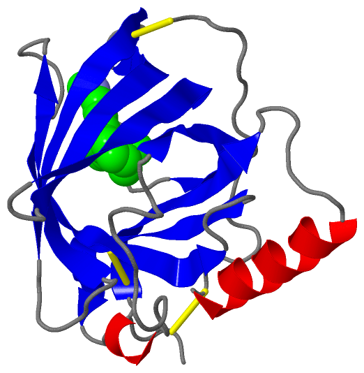
Chain A   (RET4_BOVIN | P18902)
molecular function
    GO:0016918    retinal binding    Interacting selectively and non-covalently with retinal, one of the forms of vitamin A. Retinal plays an important role in the visual process in most vertebrates, combining with opsins to form visual pigments in the retina.
    GO:0005501    retinoid binding    Interacting selectively and non-covalently with retinoids, any member of a class of isoprenoids that contain or are derived from four prenyl groups linked head-to-tail. Retinoids include retinol and retinal and structurally similar natural derivatives or synthetic compounds, but need not have vitamin A activity.
    GO:0019841    retinol binding    Interacting selectively and non-covalently with retinol, vitamin A1, 2,6,6-trimethyl-1-(9'-hydroxy-3',7'-dimethylnona-1',3',5',7'-tetraenyl)cyclohex-1-ene, one of the three components that makes up vitamin A. Retinol is an intermediate in the vision cycle and it also plays a role in growth and differentiation.
    GO:0036094    small molecule binding    Interacting selectively and non-covalently with a small molecule, any low molecular weight, monomeric, non-encoded molecule.
    GO:0005215    transporter activity    Enables the directed movement of substances (such as macromolecules, small molecules, ions) into, out of or within a cell, or between cells.
biological process
    GO:0006810    transport    The directed movement of substances (such as macromolecules, small molecules, ions) or cellular components (such as complexes and organelles) into, out of or within a cell, or between cells, or within a multicellular organism by means of some agent such as a transporter, pore or motor protein.
cellular component
    GO:0005576    extracellular region    The space external to the outermost structure of a cell. For cells without external protective or external encapsulating structures this refers to space outside of the plasma membrane. This term covers the host cell environment outside an intracellular parasite.

 Visualization

(-) Interactive Views

Asymmetric/Biological Unit
  Complete Structure
    Jena3D(integrated viewing of ligand, site, SAP, PROSITE, SCOP information)
    WebMol | AstexViewer[tm]@PDBe
(Java Applets, require no local installation except for Java; loading may be slow)
    STRAP
(Java WebStart application, automatic local installation, requires Java; full application with system access!)
    RasMol
(require local installation)
    Molscript (VRML)
(requires installation of a VRML viewer; select preferred view via VRML and generate a mono or stereo PDF format file)
 
  Ligands, Modified Residues, Ions
    ETR  [ RasMol | Jena3D ]  +environment [ RasMol | Jena3D ]
 
  Sites
    AC1  [ RasMol ]  +environment [ RasMol ]
 
  Cis Peptide Bonds
(no "Cis Peptide Bonds" information available for 1erb)
 

(-) Still Images

Jmol
  protein: cartoon or spacefill or dots and stick; nucleic acid: cartoon and stick; ligands: spacefill; active site: stick
Molscript
  protein, nucleic acid: cartoon; ligands: spacefill; active site: ball and stick

 Databases and Analysis Tools

(-) Databases

Access by PDB/NDB ID
  1erb
    Family and Domain InformationProDom | SYSTERS
    General Structural InformationGlycoscienceDB | MMDB | NDB | OCA | PDB | PDBe | PDBj | PDBsum | PDBWiki | PQS | PROTEOPEDIA
    Orientation in MembranesOPM
    Protein SurfaceSURFACE
    Secondary StructureDSSP (structure derived) | HSSP (homology derived)
    Structural GenomicsGeneCensus
    Structural NeighboursCE | VAST
    Structure ClassificationCATH | Dali | SCOP
    Validation and Original DataBMRB Data View | BMRB Restraints Grid | EDS | PROCHECK | RECOORD | WHAT_CHECK
 
Access by UniProt ID/Accession number
  RET4_BOVIN | P18902
    Comparative Protein Structure ModelsModBase
    Genomic InformationEnsembl
    Protein-protein InteractionDIP
    Sequence, Family and Domain InformationInterPro | Pfam | SMART | UniProtKB/SwissProt
 
Access by Enzyme Classificator   (EC Number)
  (no 'Enzyme Classificator' available)
    General Enzyme InformationBRENDA | EC-PDB | Enzyme | IntEnz
    PathwayKEGG | MetaCyc
 
Access by Disease Identifier   (MIM ID)
  (no 'MIM ID' available)
    Disease InformationOMIM
 
Access by GenAge ID
  (no 'GenAge ID' available)
    Age Related InformationGenAge

(-) Analysis Tools

Access by PDB/NDB ID
    Domain InformationXDom
    Interatomic Contacts of Structural UnitsCSU
    Ligand-protein ContactsLPC
    Protein CavitiescastP
    Sequence and Secondary StructurePDBCartoon
    Structure AlignmentSTRAP(Java WebStart application, automatic local installation, requires Java; full application with system access!)
    Structure and Sequence BrowserSTING
 
Access by UniProt ID/Accession number
  RET4_BOVIN | P18902
    Protein Disorder PredictionDisEMBL | FoldIndex | GLOBPLOT (for more information see DisProt)

 Related Entries

(-) Entries Sharing at Least One Protein Chain (UniProt ID)

UniProtKB/Swiss-Prot
        RET4_BOVIN | P189021fel 1fem 1fen 1hbp 1hbq 1kt3 1kt4 1kt5 1kt6 1kt7

(-) Related Entries Specified in the PDB File

(no "Related Entries Specified in the PDB File" available for 1ERB)