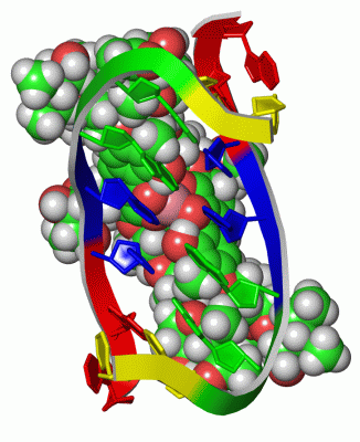
NMR Structure (13, 13)
| No. | Name | Evidence | Residues | Description |
|---|
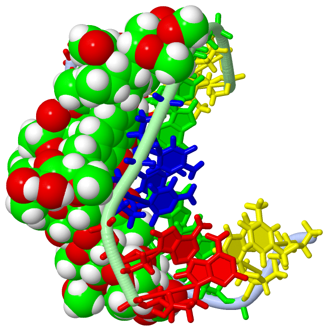
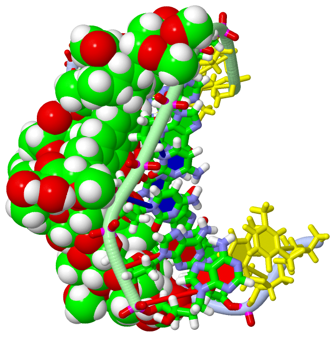
| 01 | AC1 | SOFTWARE | DG B:14 , DC B:15 , 2GL B:22 , ERI B:34 | BINDING SITE FOR RESIDUE 1GL B 21 |
| 02 | AC2 | SOFTWARE | DG B:14 , DC B:15 , 1GL B:21 , CPH B:23 , ERI B:34 , DDA B:35 | BINDING SITE FOR RESIDUE 2GL B 22 |
| 03 | AC3 | SOFTWARE | DG A:3 , 2GL A:32 , CPH A:33 , DA B:17 , DA B:18 , DDA B:25 | BINDING SITE FOR RESIDUE ERI B 24 |
| 04 | AC4 | SOFTWARE | 2GL A:32 , CPH A:33 , ERI B:24 , DDA B:26 | BINDING SITE FOR RESIDUE DDA B 25 |
| 05 | AC5 | SOFTWARE | CPH A:33 , DC B:16 , CPH B:23 , DDA B:25 | BINDING SITE FOR RESIDUE DDA B 26 |
| 06 | AC6 | SOFTWARE | DG A:4 , DC A:5 , 2GL A:32 | BINDING SITE FOR RESIDUE 1GL A 31 |
| 07 | AC7 | SOFTWARE | DG A:4 , DC A:5 , 1GL A:31 , CPH A:33 , ERI B:24 , DDA B:25 | BINDING SITE FOR RESIDUE 2GL A 32 |
| 08 | AC8 | SOFTWARE | DA A:7 , DG B:13 , DG B:14 , 1GL B:21 , 2GL B:22 , CPH B:23 , DDA B:35 | BINDING SITE FOR RESIDUE ERI B 34 |
| 09 | AC9 | SOFTWARE | 2GL B:22 , CPH B:23 , ERI B:34 , DDA B:36 | BINDING SITE FOR RESIDUE DDA B 35 |
| 10 | BC1 | SOFTWARE | CPH A:33 , DDA B:35 | BINDING SITE FOR RESIDUE DDA B 36 |
| 11 | BC2 | SOFTWARE | CPH A:33 , CPH B:23 | BINDING SITE FOR RESIDUE CO A 41 |
| 12 | BC3 | SOFTWARE | DC A:6 , CPH A:33 , CO A:41 , DG B:13 , DG B:14 , DC B:15 , DC B:16 , 2GL B:22 , DDA B:26 , ERI B:34 , DDA B:35 | BINDING SITE FOR RESIDUE CPH B 23 |
| 13 | BC4 | SOFTWARE | DG A:3 , DG A:4 , DC A:5 , DC A:6 , DA A:7 , 2GL A:32 , CO A:41 , DC B:16 , CPH B:23 , ERI B:24 , DDA B:25 , DDA B:26 , DDA B:36 | BINDING SITE FOR RESIDUE CPH A 33 |
|
 Description