|
|
|
|
 Description
|
|
Compounds
|
||||||||||||||||||||||||||||||||||||
Chains, Units
Summary Information (see also Sequences/Alignments below) |
Ligands, Modified Residues, Ions (0, 0)| (no "Ligand,Modified Residues,Ions" information available for 1EDK) |
Sites (0, 0)| (no "Site" information available for 1EDK) |
SS Bonds (0, 0)| (no "SS Bond" information available for 1EDK) |
Cis Peptide Bonds (0, 0)| (no "Cis Peptide Bond" information available for 1EDK) |
SAPs(SNPs)/Variants (0, 0)| (no "SAP(SNP)/Variant" information available for 1EDK) |
PROSITE Motifs (0, 0)| (no "PROSITE Motif" information available for 1EDK) |
Exons (0, 0)| (no "Exon" information available for 1EDK) |
Sequences/Alignments
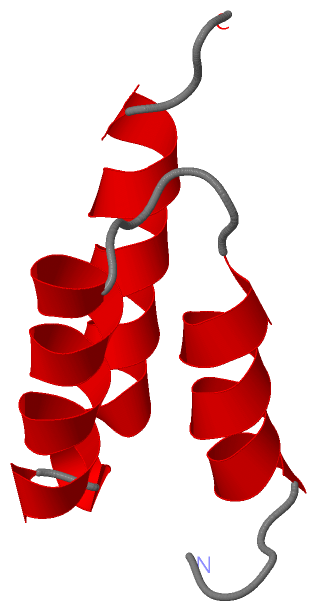
NMR StructureChain A from PDB Type:PROTEIN Length:56 aligned with SPA_STAAU | P38507 from UniProtKB/Swiss-Prot Length:508 Alignment length:56 46 56 66 76 86 SPA_STAAU 37 AQHDEAQQNAFYQVLNMPNLNADQRNGFIQSLKDDPSQSANVLGEAQKLNDSQAPK 92 SCOP domains d1edka_ A: Immunoglobulin-binding protein A modules SCOP domains CATH domains 1edkA00 A:1-56 Immunoglobulin FC, subunit C CATH domains Pfam domains -------------------------------------------------------- Pfam domains SAPs(SNPs) -------------------------------------------------------- SAPs(SNPs) PROSITE -------------------------------------------------------- PROSITE Transcript -------------------------------------------------------- Transcript 1edk A 1 AQHDEAQQNAFYQVLNMPNLNADQRNGFIQSLKDDPSQSANVLGEAQKLNDSQAPK 56 10 20 30 40 50
|
||||||||||||||||||||
SCOP Domains (1, 1)
NMR Structure
 |
CATH Domains (1, 1)
NMR Structure
|
Pfam Domains (0, 0)| (no "Pfam Domain" information available for 1EDK) |
Gene Ontology (6, 6)|
NMR Structure(hide GO term definitions) Chain A (SPA_STAAU | P38507)
|
||||||||||||||||||||||||||||||||||||||||||||||||||||||
Interactive Views
|
||||||||||||||||||||||||||||||||||||||||||||||||||||||||||||||||||||||||||||||||||||||||||||||||||||||||||||||||||||
Still Images
|
||||||||||||||||
Databases
|
||||||||||||||||||||||||||||||||||||||||||||||||||||||||||||||||||||||||||||||||||||||||||||||||||||||||||||||||||||||||||||||||||||||||||||||||||||||||||||||||
Analysis Tools
|
|||||||||||||||||||||||||||||||||||||||||||||||||||||||||||||
Entries Sharing at Least One Protein Chain (UniProt ID)
Related Entries Specified in the PDB File
|
|