|
|
|
|
 Description
|
|
Compounds
|
||||||||||||||||||||||||||||||||||||||||||||||||
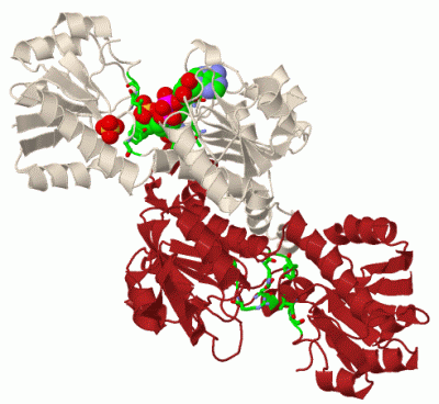
Chains, Units
Summary Information (see also Sequences/Alignments below) |
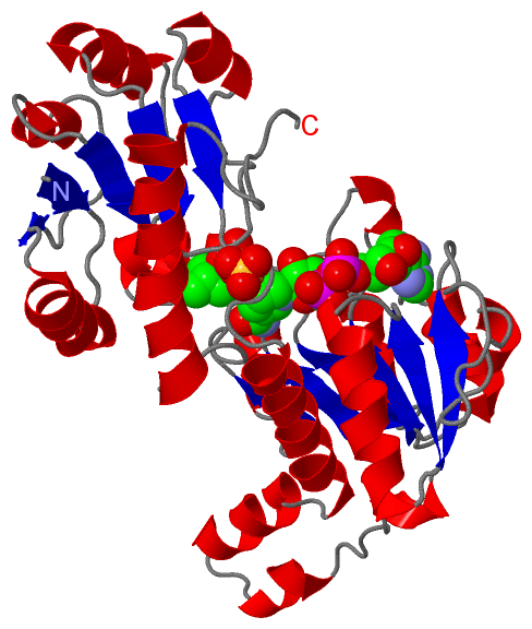
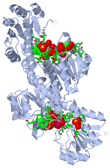
Ligands, Modified Residues, Ions (3, 4)
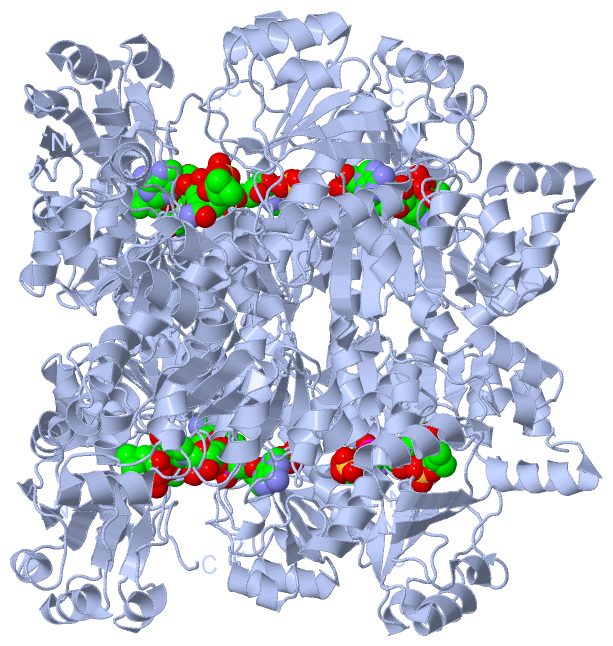
Asymmetric Unit (3, 4)
|
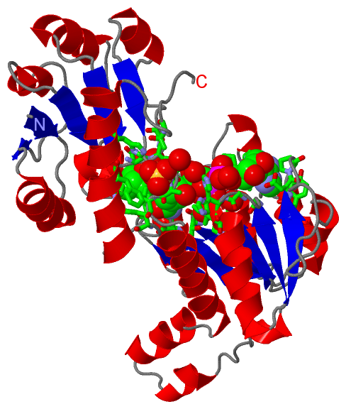
Sites (5, 5)
Asymmetric Unit (5, 5)
|
SS Bonds (0, 0)| (no "SS Bond" information available for 1DXY) |
Cis Peptide Bonds (0, 0)| (no "Cis Peptide Bond" information available for 1DXY) |
SAPs(SNPs)/Variants (0, 0)| (no "SAP(SNP)/Variant" information available for 1DXY) |
PROSITE Motifs (3, 3)
Asymmetric Unit (3, 3)
|
||||||||||||||||||||||||||||||||||||||||||||||||||||||||||||||||||||||||||||||||||||||||||||||||||||||||||||||||||||||||
Exons (0, 0)| (no "Exon" information available for 1DXY) |
Sequences/Alignments
Asymmetric UnitChain A from PDB Type:PROTEIN Length:330 aligned with DHD2_LACPA | P17584 from UniProtKB/Swiss-Prot Length:333 Alignment length:330 10 20 30 40 50 60 70 80 90 100 110 120 130 140 150 160 170 180 190 200 210 220 230 240 250 260 270 280 290 300 310 320 330 DHD2_LACPA 1 MKIIAYGARVDEIQYFKQWAKDTGNTLEYHTEFLDENTVEWAKGFDGINSLQTTPYAAGVFEKMHAYGIKFLTIRNVGTDNIDMTAMKQYGIRLSNVPAYSPAAIAEFALTDTLYLLRNMGKVQAQLQAGDYEKAGTFIGKELGQQTVGVMGTGHIGQVAIKLFKGFGAKVIAYDPYPMKGDHPDFDYVSLEDLFKQSDVIDLHVPGIEQNTHIINEAAFNLMKPGAIVINTARPNLIDTQAMLSNLKSGKLAGVGIDTYEYETEDLLNLAKHGSFKDPLWDELLGMPNVVLSPHIAYYTETAVHNMVYFSLQHLVDFLTKGETSTEVTG 330 SCOP domains d1dxya2 A:1-100,A:300-330 D-2-hydroxyisocaproate dehydrogenase d1dxya1 A:101-299 D-2-hydroxyisocaproate dehydrogenase d1dxya2 A:1-100,A:300-330 SCOP domains CATH domains 1dxyA01 A:1-101,A:299-330 NAD(P)-binding Rossmann-like Domain 1dxyA02 A:102-297 NAD(P)-binding Rossmann-like Domain -1dxyA01 A:1-101,A:299-330 CATH domains Pfam domains ------------------------------------------------------------------------------------------------------------------------------------------------------------------------------------------------------------------------------------------------------------------------------------------------------------------------------------------ Pfam domains SAPs(SNPs) ------------------------------------------------------------------------------------------------------------------------------------------------------------------------------------------------------------------------------------------------------------------------------------------------------------------------------------------ SAPs(SNPs) PROSITE ---------------------------------------------------------------------------------------------------------------------------------------------------D_2_HYDROXYACID_DH_1 ------------------D_2_HYDROXYACID_DH_2 ------D_2_HYDROXYACID_D------------------------------------------------------------------------------------------- PROSITE Transcript ------------------------------------------------------------------------------------------------------------------------------------------------------------------------------------------------------------------------------------------------------------------------------------------------------------------------------------------ Transcript 1dxy A 1 MKIIAYGARVDEIQYFKQWAKDTGNTLEYHTEFLDENTVEWAKGFDGINSLQTTPYAAGVFEKMHAYGIKFLTIRNVGTDNIDMTAMKQYGIRLSNVPAYSPAAIAEFALTDTLYLLRNMGKVQAQLQAGDYEKAGTFIGKELGQQTVGVMGTGHIGQVAIKLFKGFGAKVIAYDPYPMKGDHPDFDYVSLEDLFKQSDVIDLHVPGIEQNTHIINEAAFNLMKPGAIVINTARPNLIDTQAMLSNLKSGKLAGVGIDTYEYETEDLLNLAKHGSFKDPLWDELLGMPNVVLSPHIAYYTETAVHNMVYFSLQHLVDFLTKGETSTEVTG 330 10 20 30 40 50 60 70 80 90 100 110 120 130 140 150 160 170 180 190 200 210 220 230 240 250 260 270 280 290 300 310 320 330
|
||||||||||||||||||||
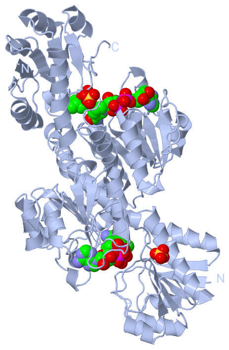
SCOP Domains (2, 2)
Asymmetric Unit
 |
CATH Domains (1, 2)
Asymmetric Unit
|
Pfam Domains (0, 0)| (no "Pfam Domain" information available for 1DXY) |
Gene Ontology (5, 5)|
Asymmetric Unit(hide GO term definitions) Chain A (DHD2_LACPA | P17584)
|
||||||||||||||||||||||||||||||||||||||||||
Interactive Views
|
|||||||||||||||||||||||||||||||||||||||||||||||||||||||||||||||||||||||||||||||||||||||||||||||||||||||||||||||||||||||||||||||||||||||||||||||||||||||||||||||||||||||||||||||||||||||
Still Images
|
||||||||||||||||
Databases
|
||||||||||||||||||||||||||||||||||||||||||||||||||||||||||||||||||||||||||||||||||||||||||||||||||||||||||||||||||||||||||||||||||||||||||||||||||||||||||||||||
Analysis Tools
|
|||||||||||||||||||||||||||||||||||||||||||||||||||||||||||||
Entries Sharing at Least One Protein Chain (UniProt ID)
Related Entries Specified in the PDB File
|
|