Show PDB file:   
         Plain Text   HTML   (compressed file size)
QuickSearch:   
by PDB,NDB,UniProt,PROSITE Code or Search Term(s)  
(-)Theoretical Model
collapse expand < >
Image Theoretical Model
Theoretical Model  (Jmol Viewer)

(-) Description

Title :  TUMOUR TARGETTING HUMAN ANTIBODIES SPECIFIC TO THE PAN-CARCINOMA ANTIGEN EPITHELIAL GLYCOPROTEIN-2 BY GUIDED SELECTION OF A MURINE ANTIBODY
 
Authors :  S. H. W. Beiboer, A. Reurs, R. C. Roovers, J. -W. Arends, N. R. J. Whitelegg, A. R. Rees, H. R. Hoogenboom
Date :  15 Dec 99  (Deposition) - 15 Dec 00  (Release) - 15 Dec 00  (Revision)
Method :  THEORETICAL MODEL
Resolution :  NOT APPLICABLE
Chains :  Theor. Model :  H,L
Keywords :  Antibody, Epg2, Tumour Targetting, Epithelial Glycoprotein 2 (Keyword Search: [Gene Ontology, PubMed, Web (Google))
 
Reference :  S. H. W. Beiboer, A. Reurs, R. C. Roovers, J. -W. Arends, N. R. J. Whitelegg, A. R. Rees, H. R. Hoogenboom
Guided Selection Of A Pan Carcinoma Specific Antibody Reveals Similar Binding Characteristics Yet Structural Divergence Between The Original Murine Antibody And Its Human Equivalent.
J. Mol. Biol. V. 296 833 2000
PubMed: search
(for further references see the PDB file header)

(-) Compounds

Molecule 1 - IMMUNOGLOBULIN VL KAPPA LIGHT CHAIN
    ChainsL
    FragmentFV RESIDUES 1-114
    Organism CommonMOUSE
    Organism ScientificMUS MUSCULUS
    Other DetailsANTI EGP-2 MOUSE ANTIBODY MOC-31
    SynonymIMMUNOGLOBULIN
 
Molecule 2 - IMMUNOGLOBULIN HEAVY CHAIN
    ChainsH
    FragmentFV RESIDUES 1-115
    Organism CommonMOUSE
    Organism ScientificMUS MUSCULUS
    Other DetailsANTI EGP-2 MOUSE ANTIBODY MOC-31
    SynonymIMMUNOGLOBULIN

 Structural Features

(-) Chains, Units

  
Theoretical Model 

Summary Information (see also Sequences/Alignments below)

(-) Ligands, Modified Residues, Ions  (0, 0)

(no "Ligand,Modified Residues,Ions" information available for 1DX2)

(-) Sites  (0, 0)

(no "Site" information available for 1DX2)

(-) SS Bonds  (2, 2)

Theoretical Model
No.Residues
1H:136 -H:210
2L:23 -L:93

(-) Cis Peptide Bonds  (2, 2)

Theoretical Model
No.Residues
1Lys L:44 -Pro L:45
2Ile L:99 -Pro L:100

 Sequence-Structure Mapping

(-) SAPs(SNPs)/Variants  (0, 0)

(no "SAP(SNP)/Variant" information available for 1DX2)

(-) PROSITE Motifs  (0, 0)

(no "PROSITE Motif" information available for 1DX2)

(-) Exons   (0, 0)

(no "Exon" information available for 1DX2)

(-) Sequences/Alignments

Theoretical Model
   Reformat: Number of residues per line =  ('0' or empty: single-line sequence representation)
  Number of residues per labelling interval =   
  UniProt sequence: complete  aligned part    
   Show mapping: SCOP domains CATH domains Pfam domains Secondary structure (by author)
SAPs(SNPs) PROSITE motifs Exons
(details for a mapped element are shown in a popup box when the mouse pointer rests over it)
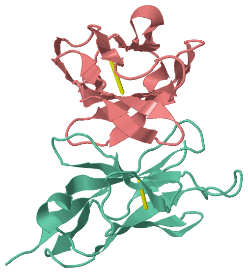
Chain H from PDB  Type:PROTEIN  Length:115
                                                                                                                                                   
               SCOP domains ------------------------------------------------------------------------------------------------------------------- SCOP domains
               CATH domains ------------------------------------------------------------------------------------------------------------------- CATH domains
               Pfam domains ------------------------------------------------------------------------------------------------------------------- Pfam domains
         Sec.struct. author ....ee...eee.....eeeeee.........eeeeeee.....eeeeeeee....eeee.hhhh..eeeeeehhh.eeeeee...hhhhheeeeeeee...eeee....eeee. Sec.struct. author
                 SAPs(SNPs) ------------------------------------------------------------------------------------------------------------------- SAPs(SNPs)
                    PROSITE ------------------------------------------------------------------------------------------------------------------- PROSITE
                 Transcript ------------------------------------------------------------------------------------------------------------------- Transcript
                 1dx2 H 115 QVKLQQSGPELKKPGETVKISCKASGYTFTNYGMNWVKQAPGKGLKWMGWINTYTGESTYADDFKGRFAFSLETSASAAYLQINNLKNEDTATYFCARFAIKGDYWGQGTTVTVS 229
                                   124       134       144       154       164       174       184       194       204       214       224     

Chain L from PDB  Type:PROTEIN  Length:114
 aligned with KV2A5_MOUSE | P03976 from UniProtKB/Swiss-Prot  Length:113

    Alignment length:114
                                                                                                                                          113 
                                    10        20        30        40        50        60        70        80        90       100       110  | 
          KV2A5_MOUSE     1 DIVMTQAVFSNPVTLGTSASISCRSSKSLLHSNGITYLYWYLQKPGQSPQLLLYQMSNLASGVPDRFSSSGSGTDFTLRISRVEAEDVGVYYCAHNLELPYTFGGGTKLEIKR-   -
               SCOP domains ------------------------------------------------------------------------------------------------------------------ SCOP domains
               CATH domains ------------------------------------------------------------------------------------------------------------------ CATH domains
               Pfam domains ------------------------------------------------------------------------------------------------------------------ Pfam domains
         Sec.struct. author ...ee....eeee.....eeeeeee............eeeeee.......eeee...ee.......eeeeee..eeeeee...hhhhheeeeeee...........eeeee... Sec.struct. author
                 SAPs(SNPs) ------------------------------------------------------------------------------------------------------------------ SAPs(SNPs)
                    PROSITE ------------------------------------------------------------------------------------------------------------------ PROSITE
                 Transcript ------------------------------------------------------------------------------------------------------------------ Transcript
                 1dx2 L   1 DIVLTQSPFSNPVTLGTSASISCRSTKSLLHSNGITYLYWYLQKPGQSPQLLIYQMSNLASGVPDRFSSSGSGTDFTLRISRVEAEDVGVYYCAQNLEIPRTFGGGTKLEIKRA 114
                                    10        20        30        40        50        60        70        80        90       100       110    

   Legend:   → Mismatch (orange background)
  - → Gap (green background, '-', border residues have a numbering label)
    → Modified Residue (blue background, lower-case, 'x' indicates undefined single-letter code, labelled with number + name)
  x → Chemical Group (purple background, 'x', labelled with number + name, e.g. ACE or NH2)
  extra numbering lines below/above indicate numbering irregularities and modified residue names etc., number ends below/above '|'

 Classification and Annotation

(-) SCOP Domains  (0, 0)

(no "SCOP Domain" information available for 1DX2)

(-) CATH Domains  (0, 0)

(no "CATH Domain" information available for 1DX2)

(-) Pfam Domains  (0, 0)

(no "Pfam Domain" information available for 1DX2)

(-) Gene Ontology  (1, 1)

Theoretical Model(hide GO term definitions)
Chain L   (KV2A5_MOUSE | P03976)
molecular function
    GO:0003823    antigen binding    Interacting selectively and non-covalently with an antigen, any substance which is capable of inducing a specific immune response and of reacting with the products of that response, the specific antibody or specifically sensitized T-lymphocytes, or both. Binding may counteract the biological activity of the antigen.

 Visualization

(-) Interactive Views

Theoretical Model
  Complete Structure
    Jena3D(integrated viewing of ligand, site, SAP, PROSITE, SCOP information)
    WebMol | AstexViewer[tm]@PDBe
(Java Applets, require no local installation except for Java; loading may be slow)
    STRAP
(Java WebStart application, automatic local installation, requires Java; full application with system access!)
    RasMol
(require local installation)
    Molscript (VRML)
(requires installation of a VRML viewer; select preferred view via VRML and generate a mono or stereo PDF format file)
 
  Ligands, Modified Residues, Ions
(no "Ligands, Modified Residues, Ions" information available for 1dx2)
 
  Sites
(no "Sites" information available for 1dx2)
 
  Cis Peptide Bonds
    Ile L:99 - Pro L:100   [ RasMol ]  
    Lys L:44 - Pro L:45   [ RasMol ]  
 

(-) Still Images

Jmol
  protein: cartoon or spacefill or dots and stick; nucleic acid: cartoon and stick; ligands: spacefill; active site: stick
Molscript
  protein, nucleic acid: cartoon; ligands: spacefill; active site: ball and stick

 Databases and Analysis Tools

(-) Databases

Access by PDB/NDB ID
  1dx2
    Family and Domain InformationProDom | SYSTERS
    General Structural InformationGlycoscienceDB | MMDB | NDB | OCA | PDB | PDBe | PDBj | PDBsum | PDBWiki | PQS | PROTEOPEDIA
    Orientation in MembranesOPM
    Protein SurfaceSURFACE
    Secondary StructureDSSP (structure derived) | HSSP (homology derived)
    Structural GenomicsGeneCensus
    Structural NeighboursCE | VAST
    Structure ClassificationCATH | Dali | SCOP
    Validation and Original DataBMRB Data View | BMRB Restraints Grid | EDS | PROCHECK | RECOORD | WHAT_CHECK
 
Access by UniProt ID/Accession number
  KV2A5_MOUSE | P03976
    Comparative Protein Structure ModelsModBase
    Genomic InformationEnsembl
    Protein-protein InteractionDIP
    Sequence, Family and Domain InformationInterPro | Pfam | SMART | UniProtKB/SwissProt
 
Access by Enzyme Classificator   (EC Number)
  (no 'Enzyme Classificator' available)
    General Enzyme InformationBRENDA | EC-PDB | Enzyme | IntEnz
    PathwayKEGG | MetaCyc
 
Access by Disease Identifier   (MIM ID)
  (no 'MIM ID' available)
    Disease InformationOMIM
 
Access by GenAge ID
  (no 'GenAge ID' available)
    Age Related InformationGenAge

(-) Analysis Tools

Access by PDB/NDB ID
    Domain InformationXDom
    Interatomic Contacts of Structural UnitsCSU
    Ligand-protein ContactsLPC
    Protein CavitiescastP
    Sequence and Secondary StructurePDBCartoon
    Structure AlignmentSTRAP(Java WebStart application, automatic local installation, requires Java; full application with system access!)
    Structure and Sequence BrowserSTING
 
Access by UniProt ID/Accession number
  KV2A5_MOUSE | P03976
    Protein Disorder PredictionDisEMBL | FoldIndex | GLOBPLOT (for more information see DisProt)

 Related Entries

(-) Entries Sharing at Least One Protein Chain (UniProt ID)

UniProtKB/Swiss-Prot
        KV2A5_MOUSE | P039763cfb 3cfc

(-) Related Entries Specified in the PDB File

1dx3 TUMOUR TARGETTING HUMAN ANTIBODIES SPECIFIC TO THE PAN-CARCINOMA ANTIGEN EPITHELIAL GLYCOPROTEIN-2 BY GUIDED SELECTION OF A MURINE ANTIBODY