|
|
|
|
 Description
|
|
Compounds
|
||||||||||||||||||||
Chains, Units
Summary Information (see also Sequences/Alignments below) |
Ligands, Modified Residues, Ions (1, 1)
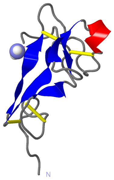
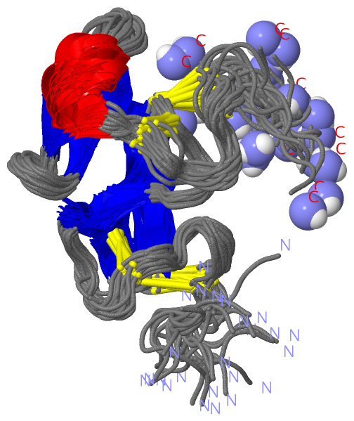
NMR Structure (1, 1)
|
Sites (1, 1)
NMR Structure (1, 1)
|
SS Bonds (5, 5)
NMR Structure
|
||||||||||||||||||||||||
Cis Peptide Bonds (0, 0)| (no "Cis Peptide Bond" information available for 1DQC) |
SAPs(SNPs)/Variants (0, 0)| (no "SAP(SNP)/Variant" information available for 1DQC) |
PROSITE Motifs (0, 0)| (no "PROSITE Motif" information available for 1DQC) |
Exons (0, 0)| (no "Exon" information available for 1DQC) |
Sequences/Alignments
NMR StructureChain A from PDB Type:PROTEIN Length:74 aligned with P91818_TACTR | P91818 from UniProtKB/TrEMBL Length:98 Alignment length:74 32 42 52 62 72 82 92 P91818_TACTR 23 YLAFRCGRYSPCLDDGPNVNLYSCCSFYNCHKCLARLENCPKGLHYNAYLKVCDWPSKAGCTSVNKECHLWKTG 96 SCOP domains d1dqca_ A: Tachycitin SCOP domains CATH domains 1dqcA00 A:1-73 Antimicrobial Protein, Tachycitin; Chain A - CATH domains Pfam domains -------------------------------------------------------------------------- Pfam domains SAPs(SNPs) -------------------------------------------------------------------------- SAPs(SNPs) PROSITE -------------------------------------------------------------------------- PROSITE Transcript -------------------------------------------------------------------------- Transcript 1dqc A 1 YLAFRCGRYSPCLDDGPNVNLYSCCSFYNCHKCLARLENCPKGLHYNAYLKVCDWPSKAGCTSVNKECHLWKTx 74 10 20 30 40 50 60 70 | 74-NH2
|
||||||||||||||||||||
SCOP Domains (1, 1)
NMR Structure
 |
CATH Domains (1, 1)
NMR Structure
|
Pfam Domains (0, 0)| (no "Pfam Domain" information available for 1DQC) |
Gene Ontology (3, 3)|
NMR Structure(hide GO term definitions) Chain A (P91818_TACTR | P91818)
|
||||||||||||||||||||||||||||||||||||
Interactive Views
|
||||||||||||||||||||||||||||||||||||||||||||||||||||||||||||||||||||||||||||||||||||||||||||||||||||||||||||||||||||||
Still Images
|
||||||||||||||||
Databases
|
||||||||||||||||||||||||||||||||||||||||||||||||||||||||||||||||||||||||||||||||||||||||||||||||||||||||||||||||||||||||||||||||||||||||||||||||||||||||||||||||
Analysis Tools
|
|||||||||||||||||||||||||||||||||||||||||||||||||||||||||||||
Entries Sharing at Least One Protein Chain (UniProt ID)
Related Entries Specified in the PDB File
|
|