|
|
|
|
 Description
|
|
Compounds
|
||||||||||||||||||||||||||||
Chains, Units
Summary Information (see also Sequences/Alignments below) |
Ligands, Modified Residues, Ions (1, 4)
Asymmetric Unit (1, 4)
|
Sites (4, 4)
Asymmetric Unit (4, 4)
|
SS Bonds (8, 8)
Asymmetric Unit
|
||||||||||||||||||||||||||||||||||||
Cis Peptide Bonds (0, 0)| (no "Cis Peptide Bond" information available for 1DKK) |
SAPs(SNPs)/Variants (0, 0)| (no "SAP(SNP)/Variant" information available for 1DKK) |
PROSITE Motifs (2, 4)
Asymmetric Unit (2, 4)
|
||||||||||||||||||||||||||||||||||||||||||||||||||||||||||||||||||||||||||||||||||||||||||||||||
Exons (0, 0)| (no "Exon" information available for 1DKK) |
Sequences/Alignments
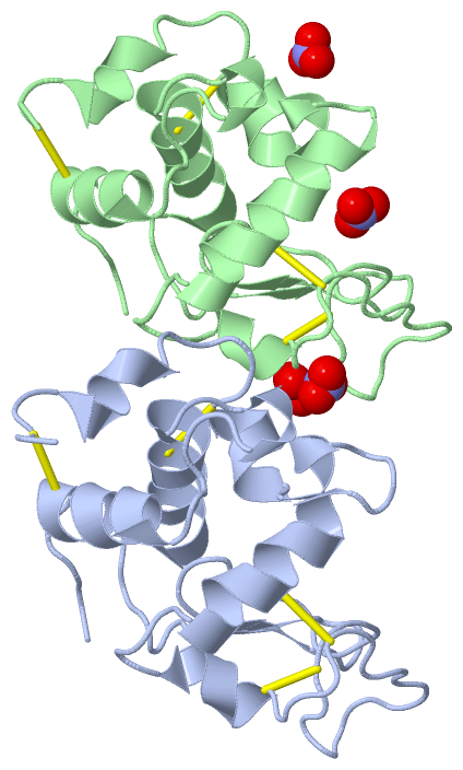
Asymmetric UnitChain A from PDB Type:PROTEIN Length:129 aligned with LYSC_COLVI | P00700 from UniProtKB/Swiss-Prot Length:129 Alignment length:129 10 20 30 40 50 60 70 80 90 100 110 120 LYSC_COLVI 1 KVFGRCELAAAMKRHGLDNYRGYSLGNWVCAAKFESNFNSQATNRNTDGSTDYGVLQINSRWWCNDGKTPGSRNLCNIPCSALLSSDITATVNCAKKIVSDGNGMNAWVAWRNRCKGTDVQAWIRGCRL 129 SCOP domains d1dkka_ A: Lysozyme SCOP domains CATH domains 1dkkA00 A:1-129 [code=1.10.530.10, no name defined] CATH domains Pfam domains --------------------------------------------------------------------------------------------------------------------------------- Pfam domains SAPs(SNPs) --------------------------------------------------------------------------------------------------------------------------------- SAPs(SNPs) PROSITE (1) LACTALBUMIN_LYSOZYME_2 PDB: A:1-129 UniProt: 1-129 PROSITE (1) PROSITE (2) ---------------------------------------------------------------------------LACTALBUMIN_LYSOZYM----------------------------------- PROSITE (2) Transcript --------------------------------------------------------------------------------------------------------------------------------- Transcript 1dkk A 1 KVFGRCELAAAMKRHGLDNYRGYSLGNWVCAAKFESNFNSQATNRNTDGSTDYGVLQINSRWWCNDGKTPGSRNLCNIPCSALLSSDITATVNCAKKIVSDGNGMNAWVAWRNRCKGTDVQAWIRGCRL 129 10 20 30 40 50 60 70 80 90 100 110 120 Chain B from PDB Type:PROTEIN Length:129 aligned with LYSC_COLVI | P00700 from UniProtKB/Swiss-Prot Length:129 Alignment length:129 10 20 30 40 50 60 70 80 90 100 110 120 LYSC_COLVI 1 KVFGRCELAAAMKRHGLDNYRGYSLGNWVCAAKFESNFNSQATNRNTDGSTDYGVLQINSRWWCNDGKTPGSRNLCNIPCSALLSSDITATVNCAKKIVSDGNGMNAWVAWRNRCKGTDVQAWIRGCRL 129 SCOP domains d1dkkb_ B: Lysozyme SCOP domains CATH domains 1dkkB00 B:1-129 [code=1.10.530.10, no name defined] CATH domains Pfam domains --------------------------------------------------------------------------------------------------------------------------------- Pfam domains SAPs(SNPs) --------------------------------------------------------------------------------------------------------------------------------- SAPs(SNPs) PROSITE (1) LACTALBUMIN_LYSOZYME_2 PDB: B:1-129 UniProt: 1-129 PROSITE (1) PROSITE (2) ---------------------------------------------------------------------------LACTALBUMIN_LYSOZYM----------------------------------- PROSITE (2) Transcript --------------------------------------------------------------------------------------------------------------------------------- Transcript 1dkk B 1 KVFGRCELAAAMKRHGLDNYRGYSLGNWVCAAKFESNFNSQATNRNTDGSTDYGVLQINSRWWCNDGKTPGSRNLCNIPCSALLSSDITATVNCAKKIVSDGNGMNAWVAWRNRCKGTDVQAWIRGCRL 129 10 20 30 40 50 60 70 80 90 100 110 120
|
||||||||||||||||||||
SCOP Domains (1, 2)
Asymmetric Unit
 |
CATH Domains (1, 2)
Asymmetric Unit
|
Pfam Domains (0, 0)| (no "Pfam Domain" information available for 1DKK) |
Gene Ontology (8, 8)|
Asymmetric Unit(hide GO term definitions) Chain A,B (LYSC_COLVI | P00700)
|
||||||||||||||||||||||||||||||||||||||||||||||||||||||||||||||||||
Interactive Views
|
||||||||||||||||||||||||||||||||||||||||||||||||||||||||||||||||||||||||||||||||||||||||||||||||||||||||||||||||||||||||||||||||||||||||||||||||||||||||||||||||||
Still Images
|
||||||||||||||||
Databases
|
||||||||||||||||||||||||||||||||||||||||||||||||||||||||||||||||||||||||||||||||||||||||||||||||||||||||||||||||||||||||||||||||||||||||||||||||||||||||||||||||
Analysis Tools
|
|||||||||||||||||||||||||||||||||||||||||||||||||||||||||||||
Entries Sharing at Least One Protein Chain (UniProt ID)
Related Entries Specified in the PDB File
|
|