Show PDB file:   
         Plain Text   HTML   (compressed file size)
QuickSearch:   
by PDB,NDB,UniProt,PROSITE Code or Search Term(s)  
(-)Asymmetric Unit
(-)Asym. Unit - sites
(-)Biological Unit 1
(-)Biol. Unit 1 - sites
(-)Biological Unit 2
collapse expand < >
Image Asymmetric Unit
Asymmetric Unit  (Jmol Viewer)
Image Asym. Unit - sites
Asym. Unit - sites  (Jmol Viewer)
Image Biological Unit 1
Biological Unit 1  (Jmol Viewer)
Image Biol. Unit 1 - sites
Biol. Unit 1 - sites  (Jmol Viewer)
Image Biological Unit 2
Biological Unit 2  (Jmol Viewer)

(-) Description

Title :  CRYSTAL STRUCTURE OF RIBOSOMAL PROTEIN L12 FROM THERMOTOGA MARITIM
 
Authors :  M. C. Wahl, G. P. Bourenkov, H. D. Bartunik, R. Huber
Date :  08 Nov 99  (Deposition) - 13 Nov 00  (Release) - 24 Feb 09  (Revision)
Method :  X-RAY DIFFRACTION
Resolution :  2.40
Chains :  Asym. Unit :  A,B,C,D
Biol. Unit 1:  A,B,C,D  (1x)
Biol. Unit 2:  A,B,C,D  (2x)
Keywords :  Dimer Formation, Flexibility, Hinge Region, Four-Helix- Bundle, Five-Helix- Bundle, Alpha-Beta Structure, Helical Hairpin, Domains, Ribosome (Keyword Search: [Gene Ontology, PubMed, Web (Google))
 
Reference :  M. C. Wahl, G. P. Bourenkov, H. D. Bartunik, R. Huber
Flexibility, Conformational Diversity And Two Dimerization Modes In Complexes Of Ribosomal Protein L12.
Embo J. V. 19 174 2000
PubMed-ID: 10637222  |  Reference-DOI: 10.1093/EMBOJ/19.2.174
(for further references see the PDB file header)

(-) Compounds

Molecule 1 - 50S RIBOSOMAL PROTEIN L7/L12
    ChainsA, B
    Organism ScientificTHERMOTOGA MARITIMA
    Organism Taxid2336
 
Molecule 2 - 50S RIBOSOMAL PROTEIN L7/L12
    ChainsC, D
    Organism ScientificTHERMOTOGA MARITIMA
    Organism Taxid2336

 Structural Features

(-) Chains, Units

  1234
Asymmetric Unit ABCD
Biological Unit 1 (1x)ABCD
Biological Unit 2 (2x)ABCD

Summary Information (see also Sequences/Alignments below)

(-) Ligands, Modified Residues, Ions  (1, 2)

Asymmetric Unit (1, 2)
No.NameCountTypeFull Name
1TBR2Ligand/IonHEXATANTALUM DODECABROMIDE
Biological Unit 1 (1, 2)
No.NameCountTypeFull Name
1TBR2Ligand/IonHEXATANTALUM DODECABROMIDE
Biological Unit 2 (1, 4)
No.NameCountTypeFull Name
1TBR4Ligand/IonHEXATANTALUM DODECABROMIDE

(-) Sites  (2, 2)

Asymmetric Unit (2, 2)
No.NameEvidenceResiduesDescription
1AC1SOFTWARELYS A:12 , LEU A:13 , ALA A:51 , GLU A:55 , LYS B:103 , GLU B:110BINDING SITE FOR RESIDUE TBR A 401
2AC2SOFTWARELYS A:103 , GLU A:110 , GLU B:11 , LYS B:12 , LEU B:13BINDING SITE FOR RESIDUE TBR A 402

(-) SS Bonds  (0, 0)

(no "SS Bond" information available for 1DD4)

(-) Cis Peptide Bonds  (0, 0)

(no "Cis Peptide Bond" information available for 1DD4)

 Sequence-Structure Mapping

(-) SAPs(SNPs)/Variants  (0, 0)

(no "SAP(SNP)/Variant" information available for 1DD4)

(-) PROSITE Motifs  (0, 0)

(no "PROSITE Motif" information available for 1DD4)

(-) Exons   (0, 0)

(no "Exon" information available for 1DD4)

(-) Sequences/Alignments

Asymmetric Unit
   Reformat: Number of residues per line =  ('0' or empty: single-line sequence representation)
  Number of residues per labelling interval =   
  UniProt sequence: complete  aligned part    
   Show mapping: SCOP domains CATH domains Pfam domains Secondary structure (by author)
SAPs(SNPs) PROSITE motifs Exons
(details for a mapped element are shown in a popup box when the mouse pointer rests over it)
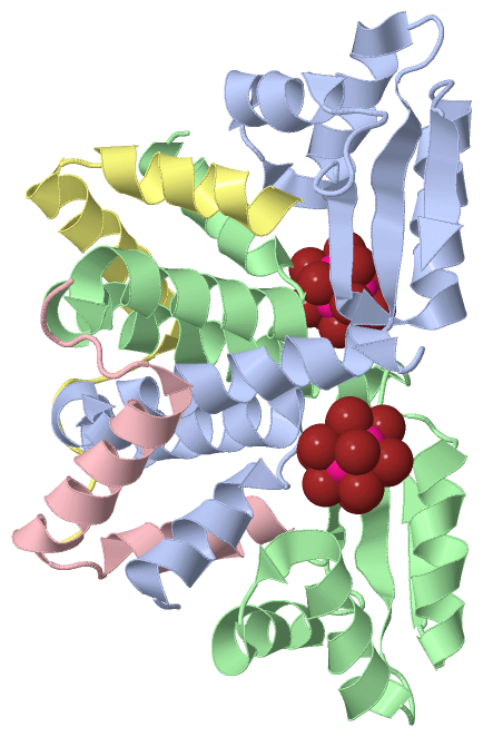
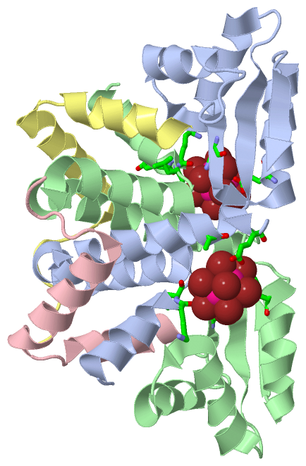
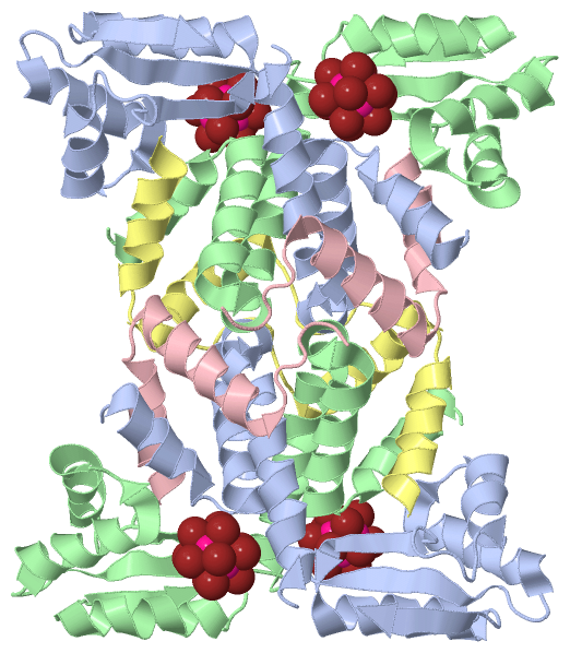
Chain A from PDB  Type:PROTEIN  Length:128
 aligned with RL7_THEMA | P29396 from UniProtKB/Swiss-Prot  Length:128

    Alignment length:128
                                    10        20        30        40        50        60        70        80        90       100       110       120        
            RL7_THEMA     1 MTIDEIIEAIEKLTVSELAELVKKLEDKFGVTAAAPVAVAAAPVAGAAAGAAQEEKTEFDVVLKSFGQNKIQVIKVVREITGLGLKEAKDLVEKAGSPDAVIKSGVSKEEAEEIKKKLEEAGAEVELK 128
               SCOP domains d1dd4a1 A:1-57                                           d1dd4a2 A:58-128 Ribosomal protein L7/12, C-terminal domain             SCOP domains
               CATH domains ---------------------------------------------------------1dd4A02 A:58-128  [code=3.30.1390.10, no name defined]                  CATH domains
               Pfam domains -------------------------------------------------------------------------------------------------------------------------------- Pfam domains
         Sec.struct. author .hhhhhhhhhh..hhhhhhhhhhhhhhhhhhhhhhhhhhhhhhhhhhhhhhhhh....eeeeeee...hhhhhhhhhhhhhh.hhhhhhhhhh.......eeeeeehhhhhhhhhhhhhh...eeee. Sec.struct. author
                 SAPs(SNPs) -------------------------------------------------------------------------------------------------------------------------------- SAPs(SNPs)
                    PROSITE -------------------------------------------------------------------------------------------------------------------------------- PROSITE
                 Transcript -------------------------------------------------------------------------------------------------------------------------------- Transcript
                 1dd4 A   1 MTIDEIIEAIEKLTVSELAELVKKLEDKFGVTAAAPVAVAAAPVAGAAAGAAQEEKTEFDVVLKSFGQNKIQVIKVVREITGLGLKEAKDLVEKAGSPDAVIKSGVSKEEAEEIKKKLEEAGAEVELK 128
                                    10        20        30        40        50        60        70        80        90       100       110       120        

Chain B from PDB  Type:PROTEIN  Length:128
 aligned with RL7_THEMA | P29396 from UniProtKB/Swiss-Prot  Length:128

    Alignment length:128
                                    10        20        30        40        50        60        70        80        90       100       110       120        
            RL7_THEMA     1 MTIDEIIEAIEKLTVSELAELVKKLEDKFGVTAAAPVAVAAAPVAGAAAGAAQEEKTEFDVVLKSFGQNKIQVIKVVREITGLGLKEAKDLVEKAGSPDAVIKSGVSKEEAEEIKKKLEEAGAEVELK 128
               SCOP domains d1dd4b1 B:1-57                                           d1dd4b2 B:58-128 Ribosomal protein L7/12, C-terminal domain             SCOP domains
               CATH domains ---------------------------------------------------------1dd4B02 B:58-128  [code=3.30.1390.10, no name defined]                  CATH domains
               Pfam domains -------------------------------------------------------------------------------------------------------------------------------- Pfam domains
         Sec.struct. author .hhhhhhhhhhh.hhhhhhhhhhhhhhhhhhhhhhhhhhhhhhhhhhhhhhhhhh...eeeeeee...hhhhhhhhhhhhhh.hhhhhhhhhh.......eeeeee..hhhhhhhhhhhhh..eeee. Sec.struct. author
                 SAPs(SNPs) -------------------------------------------------------------------------------------------------------------------------------- SAPs(SNPs)
                    PROSITE -------------------------------------------------------------------------------------------------------------------------------- PROSITE
                 Transcript -------------------------------------------------------------------------------------------------------------------------------- Transcript
                 1dd4 B   1 MTIDEIIEAIEKLTVSELAELVKKLEDKFGVTAAAPVAVAAAPVAGAAAGAAQEEKTEFDVVLKSFGQNKIQVIKVVREITGLGLKEAKDLVEKAGSPDAVIKSGVSKEEAEEIKKKLEEAGAEVELK 128
                                    10        20        30        40        50        60        70        80        90       100       110       120        

Chain C from PDB  Type:PROTEIN  Length:35
 aligned with RL7_THEMA | P29396 from UniProtKB/Swiss-Prot  Length:128

    Alignment length:35
                                    10        20        30     
            RL7_THEMA     1 MTIDEIIEAIEKLTVSELAELVKKLEDKFGVTAAA  35
               SCOP domains d1dd4c_ C:                          SCOP domains
               CATH domains ----------------------------------- CATH domains
               Pfam domains ----------------------------------- Pfam domains
         Sec.struct. author hhhhhhhhhhh..hhhhhhhhhhhhhhhh...... Sec.struct. author
                 SAPs(SNPs) ----------------------------------- SAPs(SNPs)
                    PROSITE ----------------------------------- PROSITE
                 Transcript ----------------------------------- Transcript
                 1dd4 C   1 MTIDEIIEAIEKLTVSELAELVKKLEDKFGVTAAA  35
                                    10        20        30     

Chain D from PDB  Type:PROTEIN  Length:40
 aligned with RL7_THEMA | P29396 from UniProtKB/Swiss-Prot  Length:128

    Alignment length:40
                                    10        20        30        40
            RL7_THEMA     1 MTIDEIIEAIEKLTVSELAELVKKLEDKFGVTAAAPVAVA  40
               SCOP domains d1dd4d_ D:                               SCOP domains
               CATH domains 1dd4D01 D:1-30                ---------- CATH domains
               Pfam domains ---------------------------------------- Pfam domains
         Sec.struct. author hhhhhhhhhhhh.hhhhhhhhhhhhhhhh........... Sec.struct. author
                 SAPs(SNPs) ---------------------------------------- SAPs(SNPs)
                    PROSITE ---------------------------------------- PROSITE
                 Transcript ---------------------------------------- Transcript
                 1dd4 D   1 MTIDEIIEAIEKLTVSELAELVKKLEDKFGVTAAAPVAVA  40
                                    10        20        30        40

   Legend:   → Mismatch (orange background)
  - → Gap (green background, '-', border residues have a numbering label)
    → Modified Residue (blue background, lower-case, 'x' indicates undefined single-letter code, labelled with number + name)
  x → Chemical Group (purple background, 'x', labelled with number + name, e.g. ACE or NH2)
  extra numbering lines below/above indicate numbering irregularities and modified residue names etc., number ends below/above '|'

 Classification and Annotation

(-) SCOP Domains  (2, 6)

Asymmetric Unit

(-) CATH Domains  (2, 3)

Asymmetric Unit
(-)
Class: Alpha Beta (26913)

(-) Pfam Domains  (0, 0)

(no "Pfam Domain" information available for 1DD4)

(-) Gene Ontology  (6, 6)

Asymmetric Unit(hide GO term definitions)
Chain A,B,C,D   (RL7_THEMA | P29396)
molecular function
    GO:0003735    structural constituent of ribosome    The action of a molecule that contributes to the structural integrity of the ribosome.
biological process
    GO:0006412    translation    The cellular metabolic process in which a protein is formed, using the sequence of a mature mRNA or circRNA molecule to specify the sequence of amino acids in a polypeptide chain. Translation is mediated by the ribosome, and begins with the formation of a ternary complex between aminoacylated initiator methionine tRNA, GTP, and initiation factor 2, which subsequently associates with the small subunit of the ribosome and an mRNA or circRNA. Translation ends with the release of a polypeptide chain from the ribosome.
cellular component
    GO:0022625    cytosolic large ribosomal subunit    The large subunit of a ribosome located in the cytosol.
    GO:0005622    intracellular    The living contents of a cell; the matter contained within (but not including) the plasma membrane, usually taken to exclude large vacuoles and masses of secretory or ingested material. In eukaryotes it includes the nucleus and cytoplasm.
    GO:0030529    intracellular ribonucleoprotein complex    An intracellular macromolecular complex containing both protein and RNA molecules.
    GO:0005840    ribosome    An intracellular organelle, about 200 A in diameter, consisting of RNA and protein. It is the site of protein biosynthesis resulting from translation of messenger RNA (mRNA). It consists of two subunits, one large and one small, each containing only protein and RNA. Both the ribosome and its subunits are characterized by their sedimentation coefficients, expressed in Svedberg units (symbol: S). Hence, the prokaryotic ribosome (70S) comprises a large (50S) subunit and a small (30S) subunit, while the eukaryotic ribosome (80S) comprises a large (60S) subunit and a small (40S) subunit. Two sites on the ribosomal large subunit are involved in translation, namely the aminoacyl site (A site) and peptidyl site (P site). Ribosomes from prokaryotes, eukaryotes, mitochondria, and chloroplasts have characteristically distinct ribosomal proteins.

 Visualization

(-) Interactive Views

Asymmetric Unit
  Complete Structure
    Jena3D(integrated viewing of ligand, site, SAP, PROSITE, SCOP information)
    WebMol | AstexViewer[tm]@PDBe
(Java Applets, require no local installation except for Java; loading may be slow)
    STRAP
(Java WebStart application, automatic local installation, requires Java; full application with system access!)
    RasMol
(require local installation)
    Molscript (VRML)
(requires installation of a VRML viewer; select preferred view via VRML and generate a mono or stereo PDF format file)
 
  Ligands, Modified Residues, Ions
    TBR  [ RasMol | Jena3D ]  +environment [ RasMol | Jena3D ]
 
  Sites
    AC1  [ RasMol ]  +environment [ RasMol ]
    AC2  [ RasMol ]  +environment [ RasMol ]
 
  Cis Peptide Bonds
(no "Cis Peptide Bonds" information available for 1dd4)
 
Biological Units
  Complete Structure
    Biological Unit 1  [ Jena3D ]
    Biological Unit 2  [ Jena3D ]

(-) Still Images

Jmol
  protein: cartoon or spacefill or dots and stick; nucleic acid: cartoon and stick; ligands: spacefill; active site: stick
Molscript
  protein, nucleic acid: cartoon; ligands: spacefill; active site: ball and stick

 Databases and Analysis Tools

(-) Databases

Access by PDB/NDB ID
  1dd4
    Family and Domain InformationProDom | SYSTERS
    General Structural InformationGlycoscienceDB | MMDB | NDB | OCA | PDB | PDBe | PDBj | PDBsum | PDBWiki | PQS | PROTEOPEDIA
    Orientation in MembranesOPM
    Protein SurfaceSURFACE
    Secondary StructureDSSP (structure derived) | HSSP (homology derived)
    Structural GenomicsGeneCensus
    Structural NeighboursCE | VAST
    Structure ClassificationCATH | Dali | SCOP
    Validation and Original DataBMRB Data View | BMRB Restraints Grid | EDS | PROCHECK | RECOORD | WHAT_CHECK
 
Access by UniProt ID/Accession number
  RL7_THEMA | P29396
    Comparative Protein Structure ModelsModBase
    Genomic InformationEnsembl
    Protein-protein InteractionDIP
    Sequence, Family and Domain InformationInterPro | Pfam | SMART | UniProtKB/SwissProt
 
Access by Enzyme Classificator   (EC Number)
  (no 'Enzyme Classificator' available)
    General Enzyme InformationBRENDA | EC-PDB | Enzyme | IntEnz
    PathwayKEGG | MetaCyc
 
Access by Disease Identifier   (MIM ID)
  (no 'MIM ID' available)
    Disease InformationOMIM
 
Access by GenAge ID
  (no 'GenAge ID' available)
    Age Related InformationGenAge

(-) Analysis Tools

Access by PDB/NDB ID
    Domain InformationXDom
    Interatomic Contacts of Structural UnitsCSU
    Ligand-protein ContactsLPC
    Protein CavitiescastP
    Sequence and Secondary StructurePDBCartoon
    Structure AlignmentSTRAP(Java WebStart application, automatic local installation, requires Java; full application with system access!)
    Structure and Sequence BrowserSTING
 
Access by UniProt ID/Accession number
  RL7_THEMA | P29396
    Protein Disorder PredictionDisEMBL | FoldIndex | GLOBPLOT (for more information see DisProt)

 Related Entries

(-) Entries Sharing at Least One Protein Chain (UniProt ID)

UniProtKB/Swiss-Prot
        RL7_THEMA | P293961dd3 1l1u 1zav 1zaw 1zax

(-) Related Entries Specified in the PDB File

1dd3 1DD3 CONTAINS THE SAME MOLECULE IN SPACE GROUP C2221