Show PDB file:   
         Plain Text   HTML   (compressed file size)
QuickSearch:   
by PDB,NDB,UniProt,PROSITE Code or Search Term(s)  
(-)Biol.Unit 1 - manually
(-)Asymmetric Unit
(-)Asym. Unit - sites
(-)Biological Unit 1
(-)Biol. Unit 1 - sites
collapse expand < >
Image Biol.Unit 1 - manually
Biol.Unit 1 - manually  (Jmol Viewer)
Image Asymmetric Unit
Asymmetric Unit  (Jmol Viewer)
Image Asym. Unit - sites
Asym. Unit - sites  (Jmol Viewer)
Image Biological Unit 1
Biological Unit 1  (Jmol Viewer)
Image Biol. Unit 1 - sites
Biol. Unit 1 - sites  (Jmol Viewer)

(-) Description

Title :  MECHANISTIC IMPLICATIONS AND FAMILY RELATIONSHIPS FROM THE STRUCTURE OF DETHIOBIOTIN SYNTHETASE
 
Authors :  L. Sawyer, D. Alexeev
Date :  29 Nov 94  (Deposition) - 20 Apr 95  (Release) - 24 Feb 09  (Revision)
Method :  X-RAY DIFFRACTION
Resolution :  1.80
Chains :  Asym. Unit :  A
Biol. Unit 1:  A  (2x)
Keywords :  Biotin Biosynthesis (Keyword Search: [Gene Ontology, PubMed, Web (Google))
 
Reference :  D. Alexeev, R. L. Baxter, L. Sawyer
Mechanistic Implications And Family Relationships From The Structure Of Dethiobiotin Synthetase.
Structure V. 2 1061 1994
PubMed-ID: 7881906  |  Reference-DOI: 10.1016/S0969-2126(94)00109-X
(for further references see the PDB file header)

(-) Compounds

Molecule 1 - DETHIOBIOTIN SYNTHETASE
    ChainsA
    EC Number6.3.3.3
    EngineeredYES
    Organism ScientificESCHERICHIA COLI
    Organism Taxid562

 Structural Features

(-) Chains, Units

  1
Asymmetric Unit A
Biological Unit 1 (2x)A

Summary Information (see also Sequences/Alignments below)

(-) Ligands, Modified Residues, Ions  (1, 3)

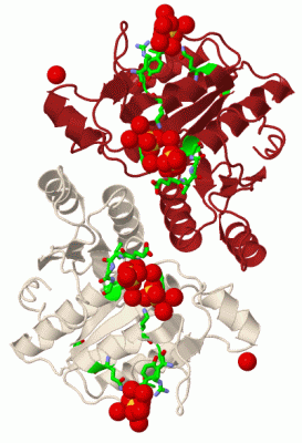
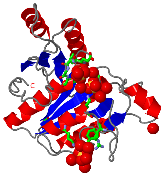
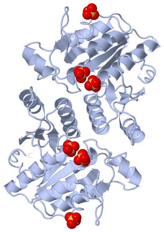
Asymmetric Unit (1, 3)
No.NameCountTypeFull Name
1SO43Ligand/IonSULFATE ION
Biological Unit 1 (1, 6)
No.NameCountTypeFull Name
1SO46Ligand/IonSULFATE ION

(-) Sites  (3, 3)

Asymmetric Unit (3, 3)
No.NameEvidenceResiduesDescription
1AC1SOFTWAREGLU A:12 , VAL A:13 , GLY A:14 , LYS A:15 , HOH A:401 , HOH A:402 , HOH A:446 , HOH A:466BINDING SITE FOR RESIDUE SO4 A 301
2AC2SOFTWARELYS A:15 , LYS A:37 , ALA A:117 , GLY A:118 , HOH A:401 , HOH A:410 , HOH A:446 , HOH A:467BINDING SITE FOR RESIDUE SO4 A 302
3AC3SOFTWARESER A:1 , ARG A:51 , GLN A:59 , TYR A:68 , HOH A:405 , HOH A:434 , HOH A:435 , HOH A:534 , HOH A:591 , HOH A:593BINDING SITE FOR RESIDUE SO4 A 303

(-) SS Bonds  (0, 0)

(no "SS Bond" information available for 1DBS)

(-) Cis Peptide Bonds  (0, 0)

(no "Cis Peptide Bond" information available for 1DBS)

 Sequence-Structure Mapping

(-) SAPs(SNPs)/Variants  (0, 0)

(no "SAP(SNP)/Variant" information available for 1DBS)

(-) PROSITE Motifs  (0, 0)

(no "PROSITE Motif" information available for 1DBS)

(-) Exons   (0, 0)

(no "Exon" information available for 1DBS)

(-) Sequences/Alignments

Asymmetric Unit
   Reformat: Number of residues per line =  ('0' or empty: single-line sequence representation)
  Number of residues per labelling interval =   
  UniProt sequence: complete  aligned part    
   Show mapping: SCOP domains CATH domains Pfam domains Secondary structure (by author)
SAPs(SNPs) PROSITE motifs Exons
(details for a mapped element are shown in a popup box when the mouse pointer rests over it)
Chain A from PDB  Type:PROTEIN  Length:224
 aligned with BIOD1_ECOLI | P13000 from UniProtKB/Swiss-Prot  Length:225

    Alignment length:224
                                    11        21        31        41        51        61        71        81        91       101       111       121       131       141       151       161       171       181       191       201       211       221    
          BIOD1_ECOLI     2 SKRYFVTGTDTEVGKTVASCALLQAAKAAGYRTAGYKPVASGSEKTPEGLRNSDALALQRNSSLQLDYATVNPYTFAEPTSPHIISAQEGRPIESLVMSAGLRALEQQADWVLVEGAGGWFTPLSDTFTFADWVTQEQLPVILVVGVKLGCINHAMLTAQVIQHAGLTLAGWVANDVTPPGKRHAEYMTTLTRMIPAPLLGEIPWLAENPENAATGKYINLALL 225
               SCOP domains d1dbsa_ A: Dethiobiotin synthetase                                                                                                                                                                                               SCOP domains
               CATH domains 1dbsA00 A:1-224 P-loop containing nucleotide triphosphate hydrolases                                                                                                                                                             CATH domains
               Pfam domains -------------------------------------------------------------------------------------------------------------------------------------------------------------------------------------------------------------------------------- Pfam domains
         Sec.struct. author .eeeeee.......hhhhhhhhhhhhhhh...eeee.eeee...........hhhhhhhhh............eee.....hhhhhhhh.....hhhhhhhhhhhh....eeeeee.............hhhhhhhh...eeeee.....hhhhhhhhhhhhhhh......eeee........hhhhhhhhhhh.....eeee..................... Sec.struct. author
                 SAPs(SNPs) -------------------------------------------------------------------------------------------------------------------------------------------------------------------------------------------------------------------------------- SAPs(SNPs)
                    PROSITE -------------------------------------------------------------------------------------------------------------------------------------------------------------------------------------------------------------------------------- PROSITE
                 Transcript -------------------------------------------------------------------------------------------------------------------------------------------------------------------------------------------------------------------------------- Transcript
                 1dbs A   1 SKRYFVTGTDTEVGKTVASCALLQAAKAAGYRTAGYKPVASGSEKTPEGLRNSDALALQRNSSLQLDYATVNPYTFAEPTSPHIISAQEGRPIESLVMSAGLRALEQQADWVLVEGAGGWFTPLSDTFTFADWVTQEQLPVILVVGVKLGCINHAMLTAQVIQHAGLTLAGWVANDVTPPGKRHAEYMTTLTRMIPAPLLGEIPWLAENPENAATGKYINLALL 224
                                    10        20        30        40        50        60        70        80        90       100       110       120       130       140       150       160       170       180       190       200       210       220    
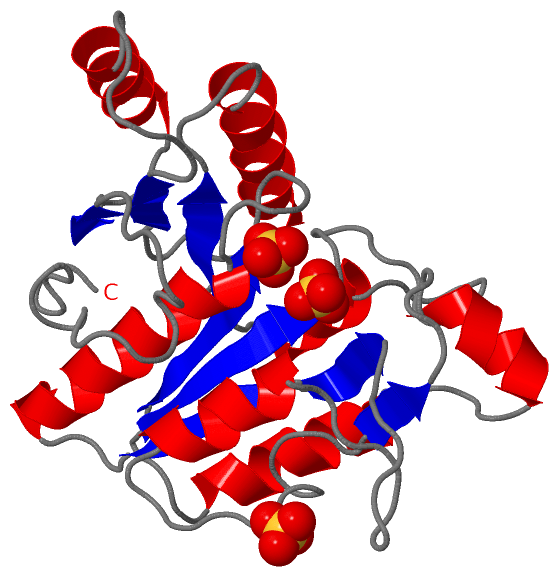
   Legend:   → Mismatch (orange background)
  - → Gap (green background, '-', border residues have a numbering label)
    → Modified Residue (blue background, lower-case, 'x' indicates undefined single-letter code, labelled with number + name)
  x → Chemical Group (purple background, 'x', labelled with number + name, e.g. ACE or NH2)
  extra numbering lines below/above indicate numbering irregularities and modified residue names etc., number ends below/above '|'

 Classification and Annotation

(-) SCOP Domains  (1, 1)

Asymmetric Unit

(-) CATH Domains  (1, 1)

Asymmetric Unit
(-)
Class: Alpha Beta (26913)

(-) Pfam Domains  (0, 0)

(no "Pfam Domain" information available for 1DBS)

(-) Gene Ontology  (9, 9)

Asymmetric Unit(hide GO term definitions)
Chain A   (BIOD1_ECOLI | P13000)
molecular function
    GO:0005524    ATP binding    Interacting selectively and non-covalently with ATP, adenosine 5'-triphosphate, a universally important coenzyme and enzyme regulator.
    GO:0004141    dethiobiotin synthase activity    Catalysis of the reaction: 7,8-diaminononanoate + ATP + CO(2) = ADP + dethiobiotin + 4 H(+) + phosphate.
    GO:0016874    ligase activity    Catalysis of the joining of two substances, or two groups within a single molecule, with the concomitant hydrolysis of the diphosphate bond in ATP or a similar triphosphate.
    GO:0000287    magnesium ion binding    Interacting selectively and non-covalently with magnesium (Mg) ions.
    GO:0046872    metal ion binding    Interacting selectively and non-covalently with any metal ion.
    GO:0000166    nucleotide binding    Interacting selectively and non-covalently with a nucleotide, any compound consisting of a nucleoside that is esterified with (ortho)phosphate or an oligophosphate at any hydroxyl group on the ribose or deoxyribose.
biological process
    GO:0009102    biotin biosynthetic process    The chemical reactions and pathways resulting in the formation of biotin, cis-tetrahydro-2-oxothieno(3,4-d)imidazoline-4-valeric acid.
cellular component
    GO:0005737    cytoplasm    All of the contents of a cell excluding the plasma membrane and nucleus, but including other subcellular structures.
    GO:0005829    cytosol    The part of the cytoplasm that does not contain organelles but which does contain other particulate matter, such as protein complexes.

 Visualization

(-) Interactive Views

Asymmetric Unit
  Complete Structure
    Jena3D(integrated viewing of ligand, site, SAP, PROSITE, SCOP information)
    WebMol | AstexViewer[tm]@PDBe
(Java Applets, require no local installation except for Java; loading may be slow)
    STRAP
(Java WebStart application, automatic local installation, requires Java; full application with system access!)
    RasMol
(require local installation)
    Molscript (VRML)
(requires installation of a VRML viewer; select preferred view via VRML and generate a mono or stereo PDF format file)
 
  Ligands, Modified Residues, Ions
    SO4  [ RasMol | Jena3D ]  +environment [ RasMol | Jena3D ]
 
  Sites
    AC1  [ RasMol ]  +environment [ RasMol ]
    AC2  [ RasMol ]  +environment [ RasMol ]
    AC3  [ RasMol ]  +environment [ RasMol ]
 
  Cis Peptide Bonds
(no "Cis Peptide Bonds" information available for 1dbs)
 
Biological Unit
  Complete Structure
    Biological Unit 1  [ Jena3D ]

(-) Still Images

Jmol
  protein: cartoon or spacefill or dots and stick; nucleic acid: cartoon and stick; ligands: spacefill; active site: stick
Molscript
  protein, nucleic acid: cartoon; ligands: spacefill; active site: ball and stick

 Databases and Analysis Tools

(-) Databases

Access by PDB/NDB ID
  1dbs
    Family and Domain InformationProDom | SYSTERS
    General Structural InformationGlycoscienceDB | MMDB | NDB | OCA | PDB | PDBe | PDBj | PDBsum | PDBWiki | PQS | PROTEOPEDIA
    Orientation in MembranesOPM
    Protein SurfaceSURFACE
    Secondary StructureDSSP (structure derived) | HSSP (homology derived)
    Structural GenomicsGeneCensus
    Structural NeighboursCE | VAST
    Structure ClassificationCATH | Dali | SCOP
    Validation and Original DataBMRB Data View | BMRB Restraints Grid | EDS | PROCHECK | RECOORD | WHAT_CHECK
 
Access by UniProt ID/Accession number
  BIOD1_ECOLI | P13000
    Comparative Protein Structure ModelsModBase
    Genomic InformationEnsembl
    Protein-protein InteractionDIP
    Sequence, Family and Domain InformationInterPro | Pfam | SMART | UniProtKB/SwissProt
 
Access by Enzyme Classificator   (EC Number)
  6.3.3.3
    General Enzyme InformationBRENDA | EC-PDB | Enzyme | IntEnz
    PathwayKEGG | MetaCyc
 
Access by Disease Identifier   (MIM ID)
  (no 'MIM ID' available)
    Disease InformationOMIM
 
Access by GenAge ID
  (no 'GenAge ID' available)
    Age Related InformationGenAge

(-) Analysis Tools

Access by PDB/NDB ID
    Domain InformationXDom
    Interatomic Contacts of Structural UnitsCSU
    Ligand-protein ContactsLPC
    Protein CavitiescastP
    Sequence and Secondary StructurePDBCartoon
    Structure AlignmentSTRAP(Java WebStart application, automatic local installation, requires Java; full application with system access!)
    Structure and Sequence BrowserSTING
 
Access by UniProt ID/Accession number
  BIOD1_ECOLI | P13000
    Protein Disorder PredictionDisEMBL | FoldIndex | GLOBPLOT (for more information see DisProt)

 Related Entries

(-) Entries Sharing at Least One Protein Chain (UniProt ID)

UniProtKB/Swiss-Prot
        BIOD1_ECOLI | P130001a82 1bs1 1byi 1dad 1dae 1daf 1dag 1dah 1dai 1dak 1dam 1dts

(-) Related Entries Specified in the PDB File

(no "Related Entries Specified in the PDB File" available for 1DBS)