Show PDB file:   
         Plain Text   HTML   (compressed file size)
QuickSearch:   
by PDB,NDB,UniProt,PROSITE Code or Search Term(s)  
(-)NMR Structure - model 1
(-)NMR Structure - model 1, sites
(-)NMR Structure - all models
collapse expand < >
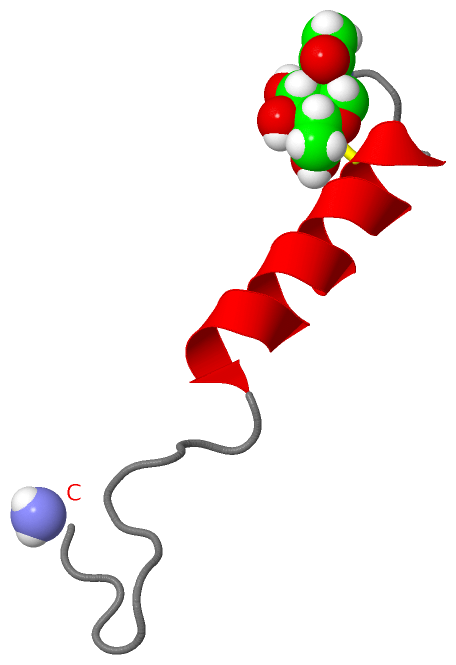
Image NMR Structure - model 1
NMR Structure - model 1  (Jmol Viewer)
Image NMR Structure - model 1, sites
NMR Structure - model 1, sites  (Jmol Viewer)
Image NMR Structure - all models
NMR Structure - all models  (Jmol Viewer)

(-) Description

Title :  GLYCOSYLATED EEL CALCITONIN
 
Authors :  Y. Hashimoto, K. Toma, J. Nishikido, K. Yamamoto, K. Haneda, T. Inazu, K. G. Valentine, S. J. Opella
Date :  16 Oct 98  (Deposition) - 28 Oct 98  (Release) - 13 Jul 11  (Revision)
Method :  SOLUTION NMR
Resolution :  NOT APPLICABLE
Chains :  NMR Structure  :  A  (10x)
NMR Structure *:  A  (1x)
Keywords :  Horomone, Calcium-Regulator, Osteoporosis, Hormone-Growth Factor Complex (Keyword Search: [Gene Ontology, PubMed, Web (Google)] )
 
Reference :  Y. Hashimoto, K. Toma, J. Nishikido, K. Yamamoto, K. Haneda, T. Inazu, K. G. Valentine, S. J. Opella
Effects Of Glycosylation On The Structure And Dynamics Of Eel Calcitonin In Micelles And Lipid Bilayers Determined By Nuclear Magnetic Resonance Spectroscopy.
Biochemistry V. 38 8377 1999
PubMed-ID: 10387083  |  Reference-DOI: 10.1021/BI983018J

(-) Compounds

Molecule 1 - PROTEIN (CALCITONIN)
    Chains: A
    Organism Common: JAPANESE EEL
    Organism Scientific: ANGUILLA JAPONICA
    Organism Taxid: 7937
    Other Details: SYNTHETICALLY GLYCOLYLATED AT ASN3

 Structural Features

(-) Chains, Units

  1
NMR Structure (10x): A
NMR Structure * (1x): A

Summary Information (see also Sequences/Alignments below)

(-) Ligands, Modified Residues, Ions  (2, 2)

NMR Structure (2, 2)
No.NameCountTypeFull Name
1NAG1Ligand/IonN-ACETYL-D-GLUCOSAMINE
2NH21Mod. Amino AcidAMINO GROUP
NMR Structure * (2, 2)
No.NameCountTypeFull Name
1NAG1Ligand/IonN-ACETYL-D-GLUCOSAMINE
2NH21Mod. Amino AcidAMINO GROUP

(-) Sites  (1, 1)

NMR Structure (1, 1)
No.NameEvidenceResiduesDescription
1AC1SOFTWARESER A:2 , ASN A:3 , THR A:6 , CYS A:7BINDING SITE FOR RESIDUE NAG A 300

(-) SS Bonds  (1, 1)

NMR Structure
No.Residues
1A:1 -A:7

(-) Cis Peptide Bonds  (0, 0)

(no "Cis Peptide Bond" information available for 1BYV)

 Sequence-Structure Mapping

(-) SAPs(SNPs)/Variants  (0, 0)

(no "SAP(SNP)/Variant" information available for 1BYV)

(-) PROSITE Motifs  (1, 1)

NMR Structure (1, 1)
 PROSITEUniProtKBPDB
No.IDACDescriptionIDLocationCountLocation
1CALCITONINPS00258 Calcitonin / CGRP / IAPP family signature.CALC_ANGJA1-16  1A:1-16
NMR Structure * (1, 1)
 PROSITEUniProtKBPDB
No.IDACDescriptionIDLocationCountLocation
1CALCITONINPS00258 Calcitonin / CGRP / IAPP family signature.CALC_ANGJA1-16  1A:1-16

(-) Exons   (0, 0)

(no "Exon" information available for 1BYV)

(-) Sequences/Alignments

NMR Structure
   Reformat: Number of residues per line =  ('0' or empty: single-line sequence representation)
  Number of residues per labelling interval =   
  UniProt sequence: complete  aligned part    
   Show mapping: SCOP domains CATH domains Pfam domains Secondary structure (by author)
SAPs(SNPs) PROSITE motifs Exons
(details for a mapped element are shown in a popup box when the mouse pointer rests over it)
Chain A from PDB  Type:PROTEIN  Length:33
 aligned with CALC_ANGJA | P01262 from UniProtKB/Swiss-Prot  Length:32

    Alignment length:33
                                                          32     
                                    10        20        30 | 
            CALC_ANGJA    1 CSNLSTCVLGKLSQELHKLQTYPRTDVGAGTP-  -
               SCOP domains d1byva_ A: Calcitonin             SCOP domains
               CATH domains --------------------------------- CATH domains
               Pfam domains --------------------------------- Pfam domains
         Sec.struct. author ....hhhhhhhhhhhhhhh.............. Sec.struct. author
                 SAPs(SNPs) --------------------------------- SAPs(SNPs)
                    PROSITE CALCITONIN      ----------------- PROSITE
                 Transcript --------------------------------- Transcript
                  1byv A  1 CSNLSTCVLGKLSQELHKLQTYPRTDVGAGTPx 33
                                    10        20        30  |
                                                           33-NH2

   Legend:   → Mismatch (orange background)
  - → Gap (green background, '-', border residues have a numbering label)
    → Modified Residue (blue background, lower-case, 'x' indicates undefined single-letter code, labelled with number + name)
  x → Chemical Group (purple background, 'x', labelled with number + name, e.g. ACE or NH2)
  extra numbering lines below/above indicate numbering irregularities and modified residue names etc., number ends below/above '|'

 Classification and Annotation

(-) SCOP Domains  (1, 1)

NMR Structure
(-)
Class: Peptides (792)

(-) CATH Domains  (0, 0)

(no "CATH Domain" information available for 1BYV)

(-) Pfam Domains  (0, 0)

(no "Pfam Domain" information available for 1BYV)

(-) Gene Ontology  (2, 2)

NMR Structure(hide GO term definitions)
Chain A   (CALC_ANGJA | P01262)
molecular function
    GO:0005179    hormone activity    The action characteristic of a hormone, any substance formed in very small amounts in one specialized organ or group of cells and carried (sometimes in the bloodstream) to another organ or group of cells in the same organism, upon which it has a specific regulatory action. The term was originally applied to agents with a stimulatory physiological action in vertebrate animals (as opposed to a chalone, which has a depressant action). Usage is now extended to regulatory compounds in lower animals and plants, and to synthetic substances having comparable effects; all bind receptors and trigger some biological process.
cellular component
    GO:0005576    extracellular region    The space external to the outermost structure of a cell. For cells without external protective or external encapsulating structures this refers to space outside of the plasma membrane. This term covers the host cell environment outside an intracellular parasite.

 Visualization

(-) Interactive Views

NMR Structure
  Complete Structure
    Jena3D(integrated viewing of ligand, site, SAP, PROSITE, SCOP information)
    WebMol | AstexViewer[tm]@PDBe
(Java Applets, require no local installation except for Java; loading may be slow)
    STRAP
(Java WebStart application, automatic local installation, requires Java; full application with system access!)
    RasMol
(require local installation)
    Molscript (VRML)
(requires installation of a VRML viewer; select preferred view via VRML and generate a mono or stereo PDF format file)
 
  Ligands, Modified Residues, Ions
    NAG  [ RasMol | Jena3D ]  +environment [ RasMol | Jena3D ]
    NH2  [ RasMol | Jena3D ]  +environment [ RasMol | Jena3D ]
 
  Sites
    AC1  [ RasMol ]  +environment [ RasMol ]
 
  Cis Peptide Bonds
(no "Cis Peptide Bonds" information available for 1byv)
 

(-) Still Images

Jmol
  protein: cartoon or spacefill or dots and stick; nucleic acid: cartoon and stick; ligands: spacefill; active site: stick
Molscript
  protein, nucleic acid: cartoon; ligands: spacefill; active site: ball and stick

 Databases and Analysis Tools

(-) Databases

Access by PDB/NDB ID
  1byv
    Family and Domain Information: ProDom | SYSTERS
    General Structural Information: GlycoscienceDB | MMDB | NDB | OCA | PDB | PDBe | PDBj | PDBsum | PDBWiki | PQS | PROTEOPEDIA
    Orientation in Membranes: OPM
    Protein Surface: SURFACE
    Secondary Structure: DSSP (structure derived) | HSSP (homology derived)
    Structural Genomics: GeneCensus
    Structural Neighbours: CE | VAST
    Structure Classification: CATH | Dali | SCOP
    Validation and Original Data: BMRB Data View | BMRB Restraints Grid | EDS | PROCHECK | RECOORD | WHAT_CHECK
 
Access by UniProt ID/Accession number
  CALC_ANGJA | P01262
    Comparative Protein Structure Models: ModBase
    Genomic Information: Ensembl
    Protein-protein Interaction: DIP
    Sequence, Family and Domain Information: InterPro | Pfam | SMART | UniProtKB/SwissProt
 
Access by Enzyme Classificator   (EC Number)
  (no 'Enzyme Classificator' available)
    General Enzyme Information: BRENDA | EC-PDB | Enzyme | IntEnz
    Pathway: KEGG | MetaCyc
 
Access by Disease Identifier   (MIM ID)
  (no 'MIM ID' available)
    Disease Information: OMIM
 
Access by GenAge ID
  (no 'GenAge ID' available)
    Age Related Information: GenAge

(-) Analysis Tools

Access by PDB/NDB ID
    Domain Information: XDom
    Interatomic Contacts of Structural Units: CSU
    Ligand-protein Contacts: LPC
    Protein Cavities: castP
    Sequence and Secondary Structure: PDBCartoon
    Structure Alignment: STRAP(Java WebStart application, automatic local installation, requires Java; full application with system access!)
    Structure and Sequence Browser: STING
 
Access by UniProt ID/Accession number
  CALC_ANGJA | P01262
    Protein Disorder Prediction: DisEMBL | FoldIndex | GLOBPLOT (for more information see DisProt)

 Related Entries

(-) Entries Sharing at Least One Protein Chain (UniProt ID)

UniProtKB/Swiss-Prot
        CALC_ANGJA | P01262: 1bku 1bzb

(-) Related Entries Specified in the PDB File

(no "Related Entries Specified in the PDB File" available for 1BYV)