|
|
|
|
 Description
|
|
Compounds
|
||||||||||||||||
Chains, Units
Summary Information (see also Sequences/Alignments below) |
Ligands, Modified Residues, Ions (9, 11)
Asymmetric Unit (9, 11)
|
Sites (11, 11)
Asymmetric Unit (11, 11)
|
SS Bonds (0, 0)| (no "SS Bond" information available for 1AX1) |
Cis Peptide Bonds (3, 3)
Asymmetric Unit
|
||||||||||||||||
SAPs(SNPs)/Variants (3, 3)
Asymmetric Unit (3, 3)
|
||||||||||||||||||||||||||||||||||||||||||||||||||||||||||||||||||||||||||||||||||||||||||||||||||||||||||||||||||||||||||||||||||||||||||||||||||||||||||||||||||||||||
PROSITE Motifs (2, 2)
Asymmetric Unit (2, 2)
|
||||||||||||||||||||||||||||||||||||||||||||||||||||||||||||||||
Exons (0, 0)| (no "Exon" information available for 1AX1) |
Sequences/Alignments
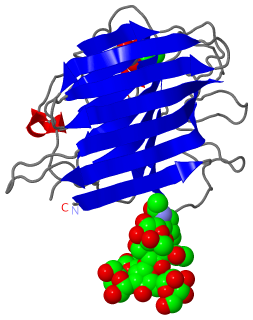
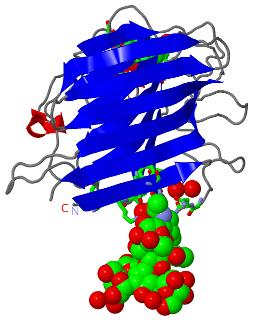
Asymmetric UnitChain A from PDB Type:PROTEIN Length:239 aligned with LEC_ERYCO | P16404 from UniProtKB/Swiss-Prot Length:281 Alignment length:239 36 46 56 66 76 86 96 106 116 126 136 146 156 166 176 186 196 206 216 226 236 246 256 LEC_ERYCO 27 VETISFSFSEFEPGNDNLTLQGAALITQSGVLQLTKINQNGMPAWDSTGRTLYAKPVHIWDMTTGTVASFETRFSFSIEQPYTRPLPADGLVFFMGPTKSKPAQGYGYLGIFNNSKQDNSYQTLGVEFDTFSNPWDPPQVPHIGIDVNSIRSIKTQPFQLDNGQVANVVIKYDASSKILHAVLVYPSSGAIYTIAEIVDVKQVLPEWVDVGLSGATGAQRDAAETHDVYSWSFQASLPE 265 SCOP domains d1ax1a_ A: Legume lectin SCOP domains CATH domains 1ax1A00 A:1-239 [code=2.60.120.200, no name defined] CATH domains Pfam domains ----------------------------------------------------------------------------------------------------------------------------------------------------------------------------------------------------------------------------------------------- Pfam domains SAPs(SNPs) ---------------------A---------------------------------------------------------------------------------------------------------------Q----------------------------------------------------------------V---------------------------------------- SAPs(SNPs) PROSITE ---------------------------------------------------------------------------------------------------------------------------LECTIN_-------------------------------------------------------------------------LECTIN_LEG-------------------------- PROSITE Transcript ----------------------------------------------------------------------------------------------------------------------------------------------------------------------------------------------------------------------------------------------- Transcript 1ax1 A 1 VETISFSFSEFEPGNDNLTLQGAALITQSGVLQLTKINQNGMPAWDSTGRTLYAKPVHIWDMTTGTVASFETRFSFSIEQPYTRPLPADGLVFFMGPTKSKPAQGYGYLGIFNNSKQDNSYQTLGVEFDTFSNPWDPPQVPHIGIDVNSIRSIKTQPFQLDNGQVANVVIKYDASSKILHAVLVYPSSGAIYTIAEIVDVKQVLPEWVDVGLSGATGAQRDAAETHDVYSWSFQASLPE 239 10 20 30 40 50 60 70 80 90 100 110 120 130 140 150 160 170 180 190 200 210 220 230
|
||||||||||||||||||||
SCOP Domains (1, 1)
Asymmetric Unit
 |
CATH Domains (1, 1)
Asymmetric Unit
|
Pfam Domains (0, 0)| (no "Pfam Domain" information available for 1AX1) |
Gene Ontology (1, 1)|
Asymmetric Unit(hide GO term definitions) Chain A (LEC_ERYCO | P16404)
|
||||||||||||
Interactive Views
|
|||||||||||||||||||||||||||||||||||||||||||||||||||||||||||||||||||||||||||||||||||||||||||||||||||||||||||||||||||||||||||||||||||||||||||||||||||||||||||||||||||||||||||||||||||||||||||||||||||||||||||||||||||||||||||||||||||||||||||||||||||||||||||||||||||||||||||||||||||||
Still Images
|
||||||||||||||||
Databases
|
||||||||||||||||||||||||||||||||||||||||||||||||||||||||||||||||||||||||||||||||||||||||||||||||||||||||||||||||||||||||||||||||||||||||||||||||||||||||||||||||
Analysis Tools
|
|||||||||||||||||||||||||||||||||||||||||||||||||||||||||||||
Entries Sharing at Least One Protein Chain (UniProt ID)
Related Entries Specified in the PDB File
|
|