| molecular function |
|---|
| | GO:0003823 | | antigen binding | | Interacting selectively and non-covalently with an antigen, any substance which is capable of inducing a specific immune response and of reacting with the products of that response, the specific antibody or specifically sensitized T-lymphocytes, or both. Binding may counteract the biological activity of the antigen. |
| | GO:0004252 | | serine-type endopeptidase activity | | Catalysis of the hydrolysis of internal, alpha-peptide bonds in a polypeptide chain by a catalytic mechanism that involves a catalytic triad consisting of a serine nucleophile that is activated by a proton relay involving an acidic residue (e.g. aspartate or glutamate) and a basic residue (usually histidine). |
| biological process |
|---|
| | GO:0038095 | | Fc-epsilon receptor signaling pathway | | A series of molecular signals initiated by the binding of the Fc portion of immunoglobulin E (IgE) to an Fc-epsilon receptor on the surface of a signal-receiving cell, and ending with regulation of a downstream cellular process, e.g. transcription. The Fc portion of an immunoglobulin is its C-terminal constant region. |
| | GO:0038096 | | Fc-gamma receptor signaling pathway involved in phagocytosis | | An Fc-gamma receptor signaling pathway that contributes to the endocytic engulfment of external particulate material by phagocytes. |
| | GO:0006956 | | complement activation | | Any process involved in the activation of any of the steps of the complement cascade, which allows for the direct killing of microbes, the disposal of immune complexes, and the regulation of other immune processes; the initial steps of complement activation involve one of three pathways, the classical pathway, the alternative pathway, and the lectin pathway, all of which lead to the terminal complement pathway. |
| | GO:0006958 | | complement activation, classical pathway | | Any process involved in the activation of any of the steps of the classical pathway of the complement cascade which allows for the direct killing of microbes, the disposal of immune complexes, and the regulation of other immune processes. |
| | GO:0006955 | | immune response | | Any immune system process that functions in the calibrated response of an organism to a potential internal or invasive threat. |
| | GO:0006508 | | proteolysis | | The hydrolysis of proteins into smaller polypeptides and/or amino acids by cleavage of their peptide bonds. |
| | GO:0006898 | | receptor-mediated endocytosis | | An endocytosis process in which cell surface receptors ensure specificity of transport. A specific receptor on the cell surface binds tightly to the extracellular macromolecule (the ligand) that it recognizes; the plasma-membrane region containing the receptor-ligand complex then undergoes endocytosis, forming a transport vesicle containing the receptor-ligand complex and excluding most other plasma-membrane proteins. Receptor-mediated endocytosis generally occurs via clathrin-coated pits and vesicles. |
| | GO:0050776 | | regulation of immune response | | Any process that modulates the frequency, rate or extent of the immune response, the immunological reaction of an organism to an immunogenic stimulus. |
| cellular component |
|---|
| | GO:0072562 | | blood microparticle | | A phospholipid microvesicle that is derived from any of several cell types, such as platelets, blood cells, endothelial cells, or others, and contains membrane receptors as well as other proteins characteristic of the parental cell. Microparticles are heterogeneous in size, and are characterized as microvesicles free of nucleic acids. |
| | GO:0070062 | | extracellular exosome | | A vesicle that is released into the extracellular region by fusion of the limiting endosomal membrane of a multivesicular body with the plasma membrane. Extracellular exosomes, also simply called exosomes, have a diameter of about 40-100 nm. |
| | GO:0005576 | | extracellular region | | The space external to the outermost structure of a cell. For cells without external protective or external encapsulating structures this refers to space outside of the plasma membrane. This term covers the host cell environment outside an intracellular parasite. |
| | GO:0005886 | | plasma membrane | | The membrane surrounding a cell that separates the cell from its external environment. It consists of a phospholipid bilayer and associated proteins. |
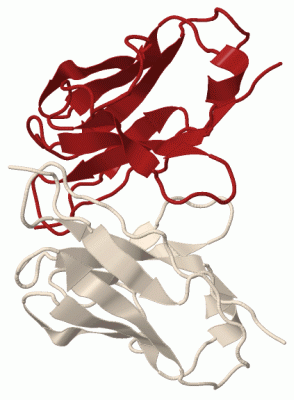
 Description
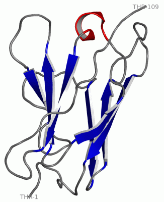