|
|
|
|
 Description
|
|
Compounds
|
||||||||||||||||||||||||
Chains, Units
Summary Information (see also Sequences/Alignments below) |
Ligands, Modified Residues, Ions (2, 9)| Asymmetric/Biological Unit (2, 9) |
Sites (10, 10)
Asymmetric Unit (10, 10)
|
SS Bonds (0, 0)| (no "SS Bond" information available for 1AKL) |
Cis Peptide Bonds (0, 0)| (no "Cis Peptide Bond" information available for 1AKL) |
SAPs(SNPs)/Variants (6, 6)
Asymmetric/Biological Unit (6, 6)
|
|||||||||||||||||||||||||||||||||||||||||||||||||||||||||||||||||||||||||||||||||||||||||||||||||||||||||||||||||||||||||||
PROSITE Motifs (2, 2)
Asymmetric/Biological Unit (2, 2)
|
||||||||||||||||||||||||||||||||
Exons (0, 0)| (no "Exon" information available for 1AKL) |
Sequences/Alignments
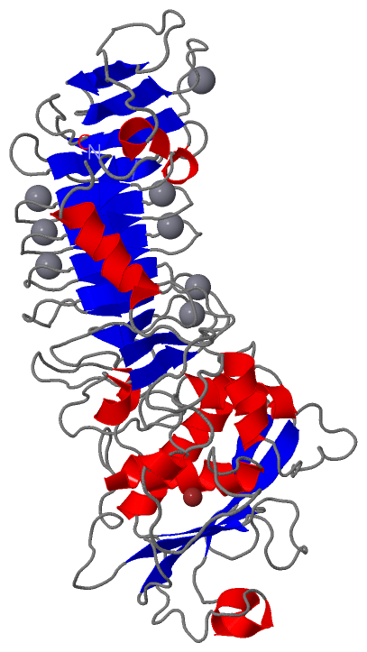
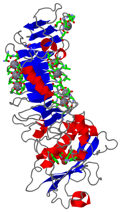
Asymmetric/Biological UnitChain A from PDB Type:PROTEIN Length:470 aligned with APRA_PSEAE | Q03023 from UniProtKB/Swiss-Prot Length:479 Alignment length:470 19 29 39 49 59 69 79 89 99 109 119 129 139 149 159 169 179 189 199 209 219 229 239 249 259 269 279 289 299 309 319 329 339 349 359 369 379 389 399 409 419 429 439 449 459 469 479 APRA_PSEAE 10 GRSDAYTQVDNFLHAYARGGDELVNGHPSYTVDQAAEQILREQASWQKAPGDSVLTLSYSFLTKPNDFFNTPWKYVSDIYSLGKFSAFSAQQQAQAKLSLQSWSDVTNIHFVDAGQGDQGDLTFGNFSSSVGGAAFAFLPDVPDALKGQSWYLINSSYSANVNPANGNYGRQTLTHEIGHTLGLSHPGDYNAGEGDPTYADATYAEDTRAYSVMSYWEEQNTGQDFKGAYSSAPLLDDIAAIQKLYGANLTTRTGDTVYGFNSNTERDFYSATSSSSKLVFSVWDAGGNDTLDFSGFSQNQKINLNEKALSDVGGLKGNVSIAAGVTVENAIGGSGSDLLIGNDVANVLKGGAGNDILYGGLGADQLWGGAGADTFVYGDIAESSAAAPDTLRDFVSGQDKIDLSGLDAFVNGGLVLQYVDAFAGKAGQAILSYDAASKAGSLAIDFSGDAHADFAINLIGQATQADIVV 479 SCOP domains d1akla2 A:1-246 Metalloprotease d1akla1 A:247-470 Metalloprotease SCOP domains CATH domains 1aklA01 1aklA02 A:18-250 Collagenase (Catalytic Domain) 1aklA01 A:1-17,A:251-470 Alkaline Protease, subunit P, domain 1 CATH domains Pfam domains -------------------------------------------------------------------------------------------------------------------------------------------------------------------------------------------------------------------------------------------------------------------------------------------------------------------------------------------------------------------------------------------------------------------------------------------------------------------------------------- Pfam domains SAPs(SNPs) ---------------------------------------------------------------------------------------------------------------------H------------------------------------------------A-----------------------------------------------------------------------------------------------------------------------------------------------------------------------------Y-------------------------------------------------------------------------------------------------------V-----R------------------L SAPs(SNPs) PROSITE ----------------------------------------------------------------------------------------------------------------------------------------------------------------------------ZINC_PROTE-----------------------------------------------------------------------------------------------------------------------------------------------------------------------------HEMOLYSIN_CALCIUM ------------------------------------------------------------------------------------------------ PROSITE Transcript -------------------------------------------------------------------------------------------------------------------------------------------------------------------------------------------------------------------------------------------------------------------------------------------------------------------------------------------------------------------------------------------------------------------------------------------------------------------------------------- Transcript 1akl A 1 GRSDAYTQVDNFLHAYARGGDELVNGHPSYTVDQAAEQILREQASWQKAPGDSVLTLSYSFLTKPNDFFNTPWKYVSDIYSLGKFSAFSAQQQAQAKLSLQSWSDVTNIHFVDAGQGDQGDLTFGNFSSSVGGAAFAFLPDVPDALKGQSWYLINSSYSANVNPANGNYGRQTLTHEIGHTLGLSHPGDYNAGEGDPTYADATYAEDTRAYSVMSYWEEQNTGQDFKGAYSSAPLLDDIAAIQKLYGANLTTRTGDTVYGFNSNTERDFYSATSSSSKLVFSVWDAGGNDTLDFSGFSQNQKINLNEKALSDVGGLKGNVSIAAGVTVENAIGGSGSDLLIGNDVANVLKGGAGNDILYGGLGADQLWGGAGADTFVYGDIAESSAAAPDTLRDFVSGQDKIDLSGLDAFVNGGLVLQYVDAFAGKAGQAILSYDAASKAGSLAIDFSGDAHADFAINLIGQATQADIVV 470 10 20 30 40 50 60 70 80 90 100 110 120 130 140 150 160 170 180 190 200 210 220 230 240 250 260 270 280 290 300 310 320 330 340 350 360 370 380 390 400 410 420 430 440 450 460 470
|
||||||||||||||||||||
SCOP Domains (2, 2)
Asymmetric/Biological Unit

|
CATH Domains (2, 2)
Asymmetric/Biological Unit
|
Pfam Domains (0, 0)| (no "Pfam Domain" information available for 1AKL) |
Gene Ontology (14, 14)|
Asymmetric/Biological Unit(hide GO term definitions) Chain A (APRA_PSEAE | Q03023)
|
||||||||||||||||||||||||||||||||||||||||||||||||||||||||||||||||||||||||||||||||||||||||||||||||||||||
Interactive Views
|
||||||||||||||||||||||||||||||||||||||||||||||||||||||||||||||||||||||||||||||||||||||||||||||||||||||||||||||||||||||||||||||||||||||||||||||||||||||||||||||||||||||||||||||||||||||||||||
Still Images
|
||||||||||||||||
Databases
|
||||||||||||||||||||||||||||||||||||||||||||||||||||||||||||||||||||||||||||||||||||||||||||||||||||||||||||||||||||||||||||||||||||||||||||||||||||||||||||||||
Analysis Tools
|
|||||||||||||||||||||||||||||||||||||||||||||||||||||||||||||
Entries Sharing at Least One Protein Chain (UniProt ID)
Related Entries Specified in the PDB File
|
|