Show PDB file:   
         Plain Text   HTML   (compressed file size)
QuickSearch:   
by PDB,NDB,UniProt,PROSITE Code or Search Term(s)  
(-)NMR Structure - manually
(-)NMR Structure - model 1
(-)NMR Structure - all models
collapse expand < >
Image NMR Structure - manually
NMR Structure - manually  (Jmol Viewer)
Image NMR Structure - model 1
NMR Structure - model 1  (Jmol Viewer)
Image NMR Structure - all models
NMR Structure - all models  (Jmol Viewer)

(-) Description

Title :  SMALL SUBUNIT C-TERMINAL INHIBITORY PEPTIDE OF MOUSE RIBONUCLEOTIDE REDUCTASE AS BOUND TO THE LARGE SUBUNIT, NMR, 26 STRUCTURES
 
Authors :  P. B. Laub, A. L. Fisher, G. T. Furst, B. A. Barwis, C. S. Hamann, B. S. Cooperman
Date :  13 Mar 97  (Deposition) - 15 May 97  (Release) - 24 Feb 09  (Revision)
Method :  SOLUTION NMR
Resolution :  NOT APPLICABLE
Chains :  NMR Structure  :  A  (26x)
Keywords :  Ribonucleotide Reductase, Peptide Inhibitors, Transferred Noesy, Restrained Molecular Dynamics, Nonstandard Type I Turn (Keyword Search: [Gene Ontology, PubMed, Web (Google))
 
Reference :  A. Fisher, P. B. Laub, B. S. Cooperman
Nmr Structure Of An Inhibitory R2 C-Terminal Peptide Bound To Mouse Ribonucleotide Reductase R1 Subunit.
Nat. Struct. Biol. V. 2 951 1995
PubMed-ID: 7583667  |  Reference-DOI: 10.1038/NSB1195-951
(for further references see the PDB file header)

(-) Compounds

Molecule 1 - RIBONUCLEOSIDE-DIPHOSPHATE REDUCTASE
    ChainsA
    EC Number1.17.4.1
    EngineeredYES
    FragmentC-TERMINAL RESIDUES AC-FTLDADF OF SMALL SUBUNIT

 Structural Features

(-) Chains, Units

  
NMR Structure (26x)

Summary Information (see also Sequences/Alignments below)

(-) Ligands, Modified Residues, Ions  (1, 1)

NMR Structure (1, 1)
No.NameCountTypeFull Name
1ACE1Mod. Amino AcidACETYL GROUP

(-) Sites  (0, 0)

(no "Site" information available for 1AFT)

(-) SS Bonds  (0, 0)

(no "SS Bond" information available for 1AFT)

(-) Cis Peptide Bonds  (0, 0)

(no "Cis Peptide Bond" information available for 1AFT)

 Sequence-Structure Mapping

(-) SAPs(SNPs)/Variants  (0, 0)

(no "SAP(SNP)/Variant" information available for 1AFT)

(-) PROSITE Motifs  (0, 0)

(no "PROSITE Motif" information available for 1AFT)

(-) Exons   (0, 0)

(no "Exon" information available for 1AFT)

(-) Sequences/Alignments

NMR Structure
   Reformat: Number of residues per line =  ('0' or empty: single-line sequence representation)
  Number of residues per labelling interval =   
  UniProt sequence: complete  aligned part    
   Show mapping: SCOP domains CATH domains Pfam domains Secondary structure (by author)
SAPs(SNPs) PROSITE motifs Exons
(details for a mapped element are shown in a popup box when the mouse pointer rests over it)
Chain A from PDB  Type:PROTEIN  Length:8
 aligned with RIR2_MOUSE | P11157 from UniProtKB/Swiss-Prot  Length:390

    Alignment length:8
           RIR2_MOUSE   383 SFTLDADF 390
               SCOP domains d1afta_  SCOP domains
               CATH domains -------- CATH domains
               Pfam domains -------- Pfam domains
         Sec.struct. author ........ Sec.struct. author
                 SAPs(SNPs) -------- SAPs(SNPs)
                    PROSITE -------- PROSITE
                 Transcript -------- Transcript
                 1aft A   0 xFTLDADF   7
                            |       
                            |       
                            0-ACE   

   Legend:   → Mismatch (orange background)
  - → Gap (green background, '-', border residues have a numbering label)
    → Modified Residue (blue background, lower-case, 'x' indicates undefined single-letter code, labelled with number + name)
  x → Chemical Group (purple background, 'x', labelled with number + name, e.g. ACE or NH2)
  extra numbering lines below/above indicate numbering irregularities and modified residue names etc., number ends below/above '|'

 Classification and Annotation

(-) SCOP Domains  (1, 1)

NMR Structure
(-)
Class: Peptides (792)

(-) CATH Domains  (0, 0)

(no "CATH Domain" information available for 1AFT)

(-) Pfam Domains  (0, 0)

(no "Pfam Domain" information available for 1AFT)

(-) Gene Ontology  (14, 14)

NMR Structure(hide GO term definitions)
Chain A   (RIR2_MOUSE | P11157)
molecular function
    GO:0046872    metal ion binding    Interacting selectively and non-covalently with any metal ion.
    GO:0016491    oxidoreductase activity    Catalysis of an oxidation-reduction (redox) reaction, a reversible chemical reaction in which the oxidation state of an atom or atoms within a molecule is altered. One substrate acts as a hydrogen or electron donor and becomes oxidized, while the other acts as hydrogen or electron acceptor and becomes reduced.
    GO:0005515    protein binding    Interacting selectively and non-covalently with any protein or protein complex (a complex of two or more proteins that may include other nonprotein molecules).
    GO:0004748    ribonucleoside-diphosphate reductase activity, thioredoxin disulfide as acceptor    Catalysis of the reaction: 2'-deoxyribonucleoside diphosphate + thioredoxin disulfide + H2O = ribonucleoside diphosphate + thioredoxin. Thioredoxin disulfide is the oxidized form of thioredoxin.
biological process
    GO:0006260    DNA replication    The cellular metabolic process in which a cell duplicates one or more molecules of DNA. DNA replication begins when specific sequences, known as origins of replication, are recognized and bound by initiation proteins, and ends when the original DNA molecule has been completely duplicated and the copies topologically separated. The unit of replication usually corresponds to the genome of the cell, an organelle, or a virus. The template for replication can either be an existing DNA molecule or RNA.
    GO:0009186    deoxyribonucleoside diphosphate metabolic process    The chemical reactions and pathways involving a deoxyribonucleoside diphosphate, a compound consisting of a nucleobase linked to a deoxyribose sugar esterified with diphosphate on the sugar.
    GO:0009263    deoxyribonucleotide biosynthetic process    The chemical reactions and pathways resulting in the formation of a deoxyribonucleotide, a compound consisting of deoxyribonucleoside (a base linked to a deoxyribose sugar) esterified with a phosphate group at either the 3' or 5'-hydroxyl group of the sugar.
    GO:0009262    deoxyribonucleotide metabolic process    The chemical reactions and pathways involving a deoxyribonucleotide, a compound consisting of deoxyribonucleoside (a base linked to a deoxyribose sugar) esterified with a phosphate group at either the 3' or 5'-hydroxyl group of the sugar.
    GO:0055114    oxidation-reduction process    A metabolic process that results in the removal or addition of one or more electrons to or from a substance, with or without the concomitant removal or addition of a proton or protons.
    GO:0051290    protein heterotetramerization    The formation of a protein heterotetramer, a macromolecular structure consisting of four noncovalently associated subunits, of which not all are identical.
    GO:0051259    protein oligomerization    The process of creating protein oligomers, compounds composed of a small number, usually between three and ten, of component monomers; protein oligomers may be composed of different or identical monomers. Oligomers may be formed by the polymerization of a number of monomers or the depolymerization of a large protein polymer.
cellular component
    GO:0005737    cytoplasm    All of the contents of a cell excluding the plasma membrane and nucleus, but including other subcellular structures.
    GO:0005634    nucleus    A membrane-bounded organelle of eukaryotic cells in which chromosomes are housed and replicated. In most cells, the nucleus contains all of the cell's chromosomes except the organellar chromosomes, and is the site of RNA synthesis and processing. In some species, or in specialized cell types, RNA metabolism or DNA replication may be absent.
    GO:0005971    ribonucleoside-diphosphate reductase complex    An enzyme complex composed of 2-4 or more subunits, which usually contains nonheme iron and requires ATP for catalysis. Catalyzes the formation of 2'-deoxyribonucleoside diphosphate from ribonucleoside diphosphate, using either thioredoxin disulfide or glutaredoxin disulfide as an acceptor.
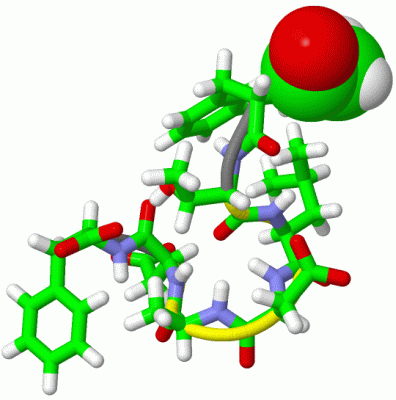
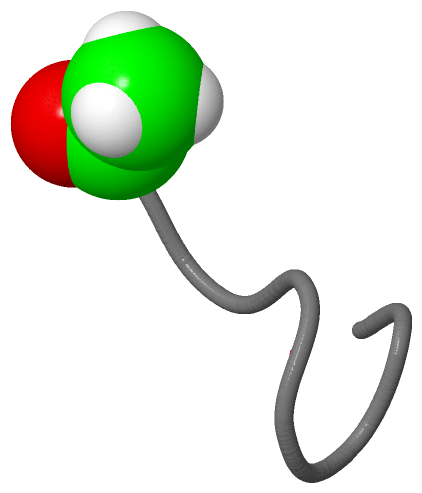
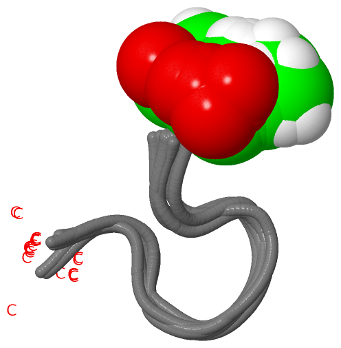
 Visualization

(-) Interactive Views

NMR Structure
  Complete Structure
    Jena3D(integrated viewing of ligand, site, SAP, PROSITE, SCOP information)
    WebMol | AstexViewer[tm]@PDBe
(Java Applets, require no local installation except for Java; loading may be slow)
    STRAP
(Java WebStart application, automatic local installation, requires Java; full application with system access!)
    RasMol
(require local installation)
    Molscript (VRML)
(requires installation of a VRML viewer; select preferred view via VRML and generate a mono or stereo PDF format file)
 
  Ligands, Modified Residues, Ions
    ACE  [ RasMol | Jena3D ]  +environment [ RasMol | Jena3D ]
 
  Sites
(no "Sites" information available for 1aft)
 
  Cis Peptide Bonds
(no "Cis Peptide Bonds" information available for 1aft)
 

(-) Still Images

Jmol
  protein: cartoon or spacefill or dots and stick; nucleic acid: cartoon and stick; ligands: spacefill; active site: stick
Molscript
  protein, nucleic acid: cartoon; ligands: spacefill; active site: ball and stick

 Databases and Analysis Tools

(-) Databases

Access by PDB/NDB ID
  1aft
    Family and Domain InformationProDom | SYSTERS
    General Structural InformationGlycoscienceDB | MMDB | NDB | OCA | PDB | PDBe | PDBj | PDBsum | PDBWiki | PQS | PROTEOPEDIA
    Orientation in MembranesOPM
    Protein SurfaceSURFACE
    Secondary StructureDSSP (structure derived) | HSSP (homology derived)
    Structural GenomicsGeneCensus
    Structural NeighboursCE | VAST
    Structure ClassificationCATH | Dali | SCOP
    Validation and Original DataBMRB Data View | BMRB Restraints Grid | EDS | PROCHECK | RECOORD | WHAT_CHECK
 
Access by UniProt ID/Accession number
  RIR2_MOUSE | P11157
    Comparative Protein Structure ModelsModBase
    Genomic InformationEnsembl
    Protein-protein InteractionDIP
    Sequence, Family and Domain InformationInterPro | Pfam | SMART | UniProtKB/SwissProt
 
Access by Enzyme Classificator   (EC Number)
  1.17.4.1
    General Enzyme InformationBRENDA | EC-PDB | Enzyme | IntEnz
    PathwayKEGG | MetaCyc
 
Access by Disease Identifier   (MIM ID)
  (no 'MIM ID' available)
    Disease InformationOMIM
 
Access by GenAge ID
  (no 'GenAge ID' available)
    Age Related InformationGenAge

(-) Analysis Tools

Access by PDB/NDB ID
    Domain InformationXDom
    Interatomic Contacts of Structural UnitsCSU
    Ligand-protein ContactsLPC
    Protein CavitiescastP
    Sequence and Secondary StructurePDBCartoon
    Structure AlignmentSTRAP(Java WebStart application, automatic local installation, requires Java; full application with system access!)
    Structure and Sequence BrowserSTING
 
Access by UniProt ID/Accession number
  RIR2_MOUSE | P11157
    Protein Disorder PredictionDisEMBL | FoldIndex | GLOBPLOT (for more information see DisProt)

 Related Entries

(-) Entries Sharing at Least One Protein Chain (UniProt ID)

UniProtKB/Swiss-Prot
        RIR2_MOUSE | P111571h0n 1h0o 1w68 1w69 1xsm

(-) Related Entries Specified in the PDB File

(no "Related Entries Specified in the PDB File" available for 1AFT)