Show PDB file:   
         Plain Text   HTML   (compressed file size)
QuickSearch:   
by PDB,NDB,UniProt,PROSITE Code or Search Term(s)  
(-)Asymmetric Unit
(-)Asym. Unit - sites
(-)Biological Unit 1
(-)Biol. Unit 1 - sites
(-)Biological Unit 2
collapse expand < >
Image Asymmetric Unit
Asymmetric Unit  (Jmol Viewer)
Image Asym. Unit - sites
Asym. Unit - sites  (Jmol Viewer)
Image Biological Unit 1
Biological Unit 1  (Jmol Viewer)
Image Biol. Unit 1 - sites
Biol. Unit 1 - sites  (Jmol Viewer)
Image Biological Unit 2
Biological Unit 2  (Jmol Viewer)

(-) Description

Title :  NOTEXIN, A PRESYNAPTIC NEUROTOXIC PHOSPHOLIPASE A2
 
Authors :  B. Westerlund, P. Nordlund, U. Uhlin, D. Eaker, H. Eklund
Date :  06 Mar 97  (Deposition) - 15 May 97  (Release) - 13 Jul 11  (Revision)
Method :  X-RAY DIFFRACTION
Resolution :  2.00
Chains :  Asym. Unit :  A
Biol. Unit 1:  A  (1x)
Biol. Unit 2:  A  (2x)
Keywords :  Hydrolase, Phospholipase A2, Lipid Degradation, Presynaptic Neurotoxin, Venom (Keyword Search: [Gene Ontology, PubMed, Web (Google))
 
Reference :  B. Westerlund, P. Nordlund, U. Uhlin, D. Eaker, H. Eklund
The Three-Dimensional Structure Of Notexin, A Presynaptic Neurotoxic Phospholipase A2 At 2. 0 A Resolution.
Febs Lett. V. 301 159 1992
PubMed-ID: 1568473  |  Reference-DOI: 10.1016/0014-5793(92)81238-H

(-) Compounds

Molecule 1 - PHOSPHOLIPASE A2
    ChainsA
    EC Number3.1.1.4
    Organism ScientificNOTECHIS SCUTATUS SCUTATUS
    Organism Taxid70142
    SecretionVENOM
    StrainSCUTATUS
    SynonymPLA2, PHOSPHATIDE SN-2 ACYLHYDROLASE

 Structural Features

(-) Chains, Units

  1
Asymmetric Unit A
Biological Unit 1 (1x)A
Biological Unit 2 (2x)A

Summary Information (see also Sequences/Alignments below)

(-) Ligands, Modified Residues, Ions  (1, 3)

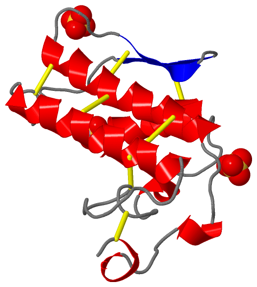
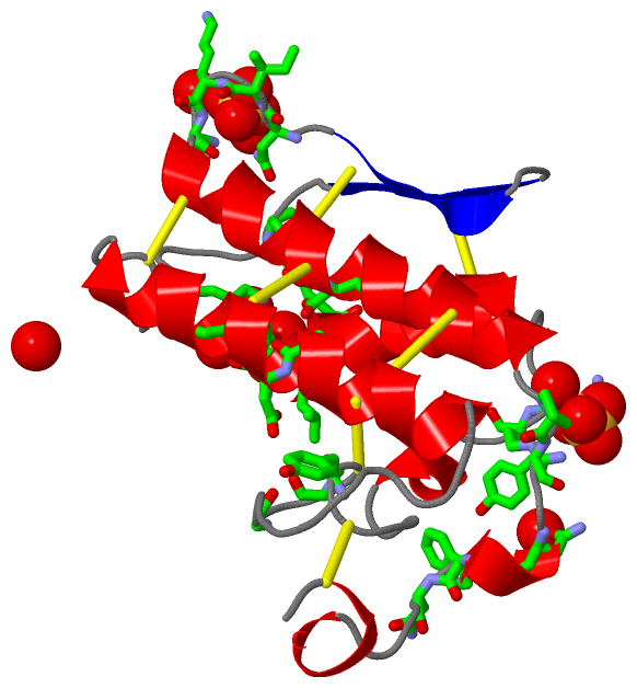
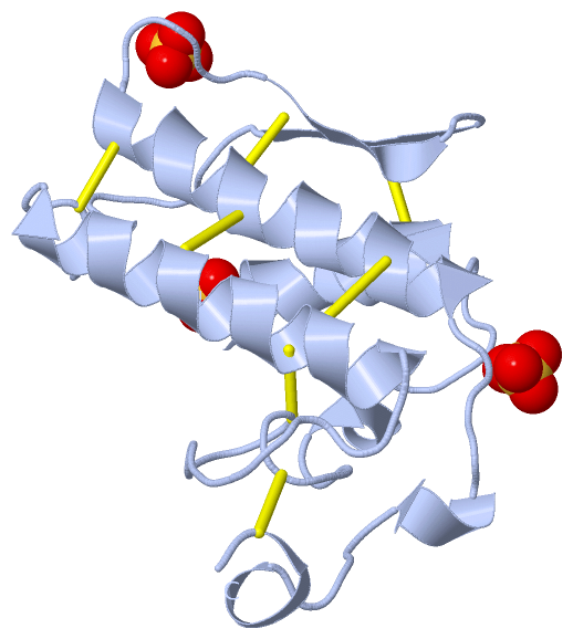
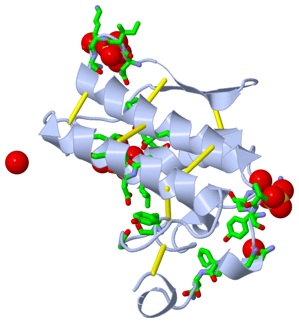
Asymmetric Unit (1, 3)
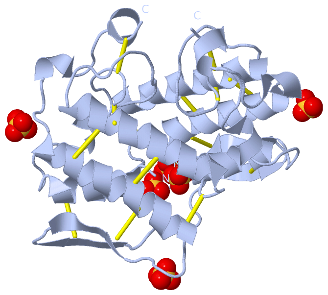
No.NameCountTypeFull Name
1SO43Ligand/IonSULFATE ION
Biological Unit 1 (1, 3)
No.NameCountTypeFull Name
1SO43Ligand/IonSULFATE ION
Biological Unit 2 (1, 6)
No.NameCountTypeFull Name
1SO46Ligand/IonSULFATE ION

(-) Sites  (5, 5)

Asymmetric Unit (5, 5)
No.NameEvidenceResiduesDescription
1AC1SOFTWAREASN A:86 , ILE A:87 , LYS A:88 , LYS A:89 , TRP A:116 , ASN A:117 , HOH A:217 , HOH A:220 , HOH A:289BINDING SITE FOR RESIDUE SO4 A 223
2AC2SOFTWAREASN A:1 , LEU A:2 , VAL A:3 , GLN A:4 , LYS A:69 , HOH A:239 , HOH A:344BINDING SITE FOR RESIDUE SO4 A 228
3AC3SOFTWARELYS A:16 , ARG A:17 , PRO A:110 , TYR A:111 , ASN A:113 , HOH A:202 , HOH A:269BINDING SITE FOR RESIDUE SO4 A 350
4CALUNKNOWNTYR A:28 , GLY A:30 , GLY A:32 , ASP A:49SITE *CAL* COMPRISES THOSE RESIDUES FORMING THE CA2+ BINDING SITE. IN THE PRESENT CA-FREE FORM THIS SITE IS EMPTY.
5CNRUNKNOWNHIS A:48 , TYR A:52 , TYR A:73 , ASP A:99SITE *CNR* COMPRISES THOSE RESIDUES FORMING THE CATALYTIC NETWORK.

(-) SS Bonds  (7, 7)

Asymmetric Unit
No.Residues
1A:11 -A:77
2A:27 -A:124
3A:29 -A:45
4A:44 -A:105
5A:51 -A:98
6A:61 -A:91
7A:84 -A:96

(-) Cis Peptide Bonds  (1, 1)

Asymmetric Unit
No.Residues
1Phe A:67 -Pro A:68

 Sequence-Structure Mapping

(-) SAPs(SNPs)/Variants  (1, 1)

Asymmetric Unit (1, 1)
  dbSNPPDB
No.SourceVariant IDVariantUniProt IDStatusIDChainVariant
1UniProtVAR_PA2B_NOTSC_001 *K16RPA2B_NOTSC  ---  ---AK16R
   * ID not provided by source

  SNP/SAP Summary Statistics (UniProtKB/Swiss-Prot)
Biological Unit 1 (1, 1)
  dbSNPPDB
No.SourceVariant IDVariantUniProt IDStatusIDChainVariant
1UniProtVAR_PA2B_NOTSC_001 *K16RPA2B_NOTSC  ---  ---AK16R
   * ID not provided by source

  SNP/SAP Summary Statistics (UniProtKB/Swiss-Prot)
Biological Unit 2 (1, 2)
  dbSNPPDB
No.SourceVariant IDVariantUniProt IDStatusIDChainVariant
1UniProtVAR_PA2B_NOTSC_001 *K16RPA2B_NOTSC  ---  ---AK16R
   * ID not provided by source

  SNP/SAP Summary Statistics (UniProtKB/Swiss-Prot)

(-) PROSITE Motifs  (2, 2)

Asymmetric Unit (2, 2)
 PROSITEUniProtKBPDB
No.IDACDescriptionIDLocationCountLocation
1PA2_HISPS00118 Phospholipase A2 histidine active site.PA2B_NOTSC44-51  1A:44-51
2PA2_ASPPS00119 Phospholipase A2 aspartic acid active site.PA2B_NOTSC89-99  1A:95-105
Biological Unit 1 (2, 2)
 PROSITEUniProtKBPDB
No.IDACDescriptionIDLocationCountLocation
1PA2_HISPS00118 Phospholipase A2 histidine active site.PA2B_NOTSC44-51  1A:44-51
2PA2_ASPPS00119 Phospholipase A2 aspartic acid active site.PA2B_NOTSC89-99  1A:95-105
Biological Unit 2 (2, 4)
 PROSITEUniProtKBPDB
No.IDACDescriptionIDLocationCountLocation
1PA2_HISPS00118 Phospholipase A2 histidine active site.PA2B_NOTSC44-51  2A:44-51
2PA2_ASPPS00119 Phospholipase A2 aspartic acid active site.PA2B_NOTSC89-99  2A:95-105

(-) Exons   (0, 0)

(no "Exon" information available for 1AE7)

(-) Sequences/Alignments

Asymmetric Unit
   Reformat: Number of residues per line =  ('0' or empty: single-line sequence representation)
  Number of residues per labelling interval =   
  UniProt sequence: complete  aligned part    
   Show mapping: SCOP domains CATH domains Pfam domains Secondary structure (by author)
SAPs(SNPs) PROSITE motifs Exons
(details for a mapped element are shown in a popup box when the mouse pointer rests over it)
Chain A from PDB  Type:PROTEIN  Length:119
 aligned with PA2B_NOTSC | P00608 from UniProtKB/Swiss-Prot  Length:119

    Alignment length:119
                                    10        20        30        40        50        60        70        80        90       100       110         
           PA2B_NOTSC     1 NLVQFSYLIQCANHGKRPTWHYMDYGCYCGAGGSGTPVDELDRCCKIHDDCYDEAGKKGCFPKMSAYDYYCGENGPYCRNIKKKCLRFVCDCDVEAAFCFAKAPYNNANWNIDTKKRCQ 119
               SCOP domains d1ae7a_ A: Snake phospholipase A2                                                                                       SCOP domains
               CATH domains 1ae7A00 A:1-125 Phospholipase A2                                                                                        CATH domains
               Pfam domains ----------------------------------------------------------------------------------------------------------------------- Pfam domains
         Sec.struct. author .hhhhhhhhhhh......hhhh.................hhhhhhhhhhhhhhhhhh...........eeee..eeee.....hhhhhhhhhhhhhhhhhhh....hhh....hhhh.. Sec.struct. author
                 SAPs(SNPs) ---------------R------------------------------------------------------------------------------------------------------- SAPs(SNPs)
                    PROSITE -------------------------------------------PA2_HIS -------------------------------------PA2_ASP    -------------------- PROSITE
                 Transcript ----------------------------------------------------------------------------------------------------------------------- Transcript
                 1ae7 A   1 NLVQFSYLIQCANHGKRPTWHYMDYGCYCGAGGSGTPVDELDRCCKIHDDCYDEAGKKGCFPKMSAYDYYCGENGPYCRNIKKKCLRFVCDCDVEAAFCFAKAPYNNANWNIDTKKRCQ 125
                                    10        20        30        40        50      ||61|       76        86        96       106       116         
                                                                                   57| ||                                                          
                                                                                    59 ||                                                          
                                                                                      61|                                                          
                                                                                       67                                                          

   Legend:   → Mismatch (orange background)
  - → Gap (green background, '-', border residues have a numbering label)
    → Modified Residue (blue background, lower-case, 'x' indicates undefined single-letter code, labelled with number + name)
  x → Chemical Group (purple background, 'x', labelled with number + name, e.g. ACE or NH2)
  extra numbering lines below/above indicate numbering irregularities and modified residue names etc., number ends below/above '|'

 Classification and Annotation

(-) SCOP Domains  (1, 1)

Asymmetric Unit

(-) CATH Domains  (1, 1)

Asymmetric Unit

(-) Pfam Domains  (0, 0)

(no "Pfam Domain" information available for 1AE7)

(-) Gene Ontology  (8, 8)

Asymmetric Unit(hide GO term definitions)
Chain A   (PA2B_NOTSC | P00608)
molecular function
    GO:0005509    calcium ion binding    Interacting selectively and non-covalently with calcium ions (Ca2+).
    GO:0016787    hydrolase activity    Catalysis of the hydrolysis of various bonds, e.g. C-O, C-N, C-C, phosphoric anhydride bonds, etc. Hydrolase is the systematic name for any enzyme of EC class 3.
    GO:0046872    metal ion binding    Interacting selectively and non-covalently with any metal ion.
    GO:0004623    phospholipase A2 activity    Catalysis of the reaction: phosphatidylcholine + H2O = 1-acylglycerophosphocholine + a carboxylate.
biological process
    GO:0016042    lipid catabolic process    The chemical reactions and pathways resulting in the breakdown of lipids, compounds soluble in an organic solvent but not, or sparingly, in an aqueous solvent.
    GO:0006629    lipid metabolic process    The chemical reactions and pathways involving lipids, compounds soluble in an organic solvent but not, or sparingly, in an aqueous solvent. Includes fatty acids; neutral fats, other fatty-acid esters, and soaps; long-chain (fatty) alcohols and waxes; sphingoids and other long-chain bases; glycolipids, phospholipids and sphingolipids; and carotenes, polyprenols, sterols, terpenes and other isoprenoids.
cellular component
    GO:0005576    extracellular region    The space external to the outermost structure of a cell. For cells without external protective or external encapsulating structures this refers to space outside of the plasma membrane. This term covers the host cell environment outside an intracellular parasite.
    GO:0072556    other organism presynaptic membrane    A presynaptic membrane that is part of another organism, i.e. a secondary organism with which the first organism is interacting. A presynaptic membrane is specialized area of membrane of the axon terminal that faces the plasma membrane of the neuron or muscle fiber with which the axon terminal establishes a synaptic junction; many synaptic junctions exhibit structural presynaptic characteristics, such as conical, electron-dense internal protrusions, that distinguish it from the remainder of the axon plasma membrane.

 Visualization

(-) Interactive Views

Asymmetric Unit
  Complete Structure
    Jena3D(integrated viewing of ligand, site, SAP, PROSITE, SCOP information)
    WebMol | AstexViewer[tm]@PDBe
(Java Applets, require no local installation except for Java; loading may be slow)
    STRAP
(Java WebStart application, automatic local installation, requires Java; full application with system access!)
    RasMol
(require local installation)
    Molscript (VRML)
(requires installation of a VRML viewer; select preferred view via VRML and generate a mono or stereo PDF format file)
 
  Ligands, Modified Residues, Ions
    SO4  [ RasMol | Jena3D ]  +environment [ RasMol | Jena3D ]
 
  Sites
    AC1  [ RasMol ]  +environment [ RasMol ]
    AC2  [ RasMol ]  +environment [ RasMol ]
    AC3  [ RasMol ]  +environment [ RasMol ]
    CAL  [ RasMol ]  +environment [ RasMol ]
    CNR  [ RasMol ]  +environment [ RasMol ]
 
  Cis Peptide Bonds
    Phe A:67 - Pro A:68   [ RasMol ]  
 
Biological Units
  Complete Structure
    Biological Unit 1  [ Jena3D ]
    Biological Unit 2  [ Jena3D ]

(-) Still Images

Jmol
  protein: cartoon or spacefill or dots and stick; nucleic acid: cartoon and stick; ligands: spacefill; active site: stick
Molscript
  protein, nucleic acid: cartoon; ligands: spacefill; active site: ball and stick

 Databases and Analysis Tools

(-) Databases

Access by PDB/NDB ID
  1ae7
    Family and Domain InformationProDom | SYSTERS
    General Structural InformationGlycoscienceDB | MMDB | NDB | OCA | PDB | PDBe | PDBj | PDBsum | PDBWiki | PQS | PROTEOPEDIA
    Orientation in MembranesOPM
    Protein SurfaceSURFACE
    Secondary StructureDSSP (structure derived) | HSSP (homology derived)
    Structural GenomicsGeneCensus
    Structural NeighboursCE | VAST
    Structure ClassificationCATH | Dali | SCOP
    Validation and Original DataBMRB Data View | BMRB Restraints Grid | EDS | PROCHECK | RECOORD | WHAT_CHECK
 
Access by UniProt ID/Accession number
  PA2B_NOTSC | P00608
    Comparative Protein Structure ModelsModBase
    Genomic InformationEnsembl
    Protein-protein InteractionDIP
    Sequence, Family and Domain InformationInterPro | Pfam | SMART | UniProtKB/SwissProt
 
Access by Enzyme Classificator   (EC Number)
  3.1.1.4
    General Enzyme InformationBRENDA | EC-PDB | Enzyme | IntEnz
    PathwayKEGG | MetaCyc
 
Access by Disease Identifier   (MIM ID)
  (no 'MIM ID' available)
    Disease InformationOMIM
 
Access by GenAge ID
  (no 'GenAge ID' available)
    Age Related InformationGenAge

(-) Analysis Tools

Access by PDB/NDB ID
    Domain InformationXDom
    Interatomic Contacts of Structural UnitsCSU
    Ligand-protein ContactsLPC
    Protein CavitiescastP
    Sequence and Secondary StructurePDBCartoon
    Structure AlignmentSTRAP(Java WebStart application, automatic local installation, requires Java; full application with system access!)
    Structure and Sequence BrowserSTING
 
Access by UniProt ID/Accession number
  PA2B_NOTSC | P00608
    Protein Disorder PredictionDisEMBL | FoldIndex | GLOBPLOT (for more information see DisProt)

 Related Entries

(-) Entries Sharing at Least One Protein Chain (UniProt ID)

UniProtKB/Swiss-Prot
        PA2B_NOTSC | P006084e4c

(-) Related Entries Specified in the PDB File

(no "Related Entries Specified in the PDB File" available for 1AE7)