Show PDB file:   
         Plain Text   HTML   (compressed file size)
QuickSearch:   
by PDB,NDB,UniProt,PROSITE Code or Search Term(s)  
(-)Asymmetric Unit
(-)Biological Unit 1
(-)Biological Unit 2
(-)Biological Unit 3
collapse expand < >
Image Asymmetric Unit
Asymmetric Unit  (Jmol Viewer)
Image Biological Unit 1
Biological Unit 1  (Jmol Viewer)
Image Biological Unit 2
Biological Unit 2  (Jmol Viewer)
Image Biological Unit 3
Biological Unit 3  (Jmol Viewer)

(-) Description

Title :  OLIGOMERIZATION DOMAIN OF HEPATITIS DELTA ANTIGEN
 
Authors :  H. J. Zuccola, J. M. Hogle
Date :  15 Apr 98  (Deposition) - 08 Jun 99  (Release) - 13 Jul 11  (Revision)
Method :  X-RAY DIFFRACTION
Resolution :  1.80
Chains :  Asym. Unit :  A,B,C,D
Biol. Unit 1:  A (2x),B (2x),C (2x),D (2x)
Biol. Unit 2:  A,C  (1x)
Biol. Unit 3:  B,D  (1x)
Keywords :  Leucine Zipper, Coiled-Coil, Oligomerization, Hepatitis Delta, Hdv (Keyword Search: [Gene Ontology, PubMed, Web (Google))
 
Reference :  H. J. Zuccola, J. E. Rozzelle, S. M. Lemon, B. W. Erickson, J. M. Hogle
Structural Basis Of The Oligomerization Of Hepatitis Delta Antigen.
Structure V. 6 821 1998
PubMed-ID: 9687364  |  Reference-DOI: 10.1016/S0969-2126(98)00084-7
(for further references see the PDB file header)

(-) Compounds

Molecule 1 - DELTA ANTIGEN
    ChainsA, B, C, D
    EngineeredYES
    FragmentOLIGOMERIZATION DOMAIN
    MutationYES
    Organism ScientificHEPATITIS DELTA VIRUS
    Organism Taxid12475
    SynonymHEPATITIS DELTA ANTIGEN

 Structural Features

(-) Chains, Units

  1234
Asymmetric Unit ABCD
Biological Unit 1 (2x)A (2x)B (2x)C (2x)D (2x)
Biological Unit 2 (1x)A C 
Biological Unit 3 (1x) B D

Summary Information (see also Sequences/Alignments below)

(-) Ligands, Modified Residues, Ions  (0, 0)

(no "Ligand,Modified Residues,Ions" information available for 1A92)

(-) Sites  (0, 0)

(no "Site" information available for 1A92)

(-) SS Bonds  (0, 0)

(no "SS Bond" information available for 1A92)

(-) Cis Peptide Bonds  (0, 0)

(no "Cis Peptide Bond" information available for 1A92)

 Sequence-Structure Mapping

(-) SAPs(SNPs)/Variants  (0, 0)

(no "SAP(SNP)/Variant" information available for 1A92)

(-) PROSITE Motifs  (0, 0)

(no "PROSITE Motif" information available for 1A92)

(-) Exons   (0, 0)

(no "Exon" information available for 1A92)

(-) Sequences/Alignments

Asymmetric Unit
   Reformat: Number of residues per line =  ('0' or empty: single-line sequence representation)
  Number of residues per labelling interval =   
  UniProt sequence: complete  aligned part    
   Show mapping: SCOP domains CATH domains Pfam domains Secondary structure (by author)
SAPs(SNPs) PROSITE motifs Exons
(details for a mapped element are shown in a popup box when the mouse pointer rests over it)
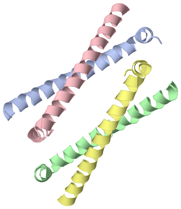
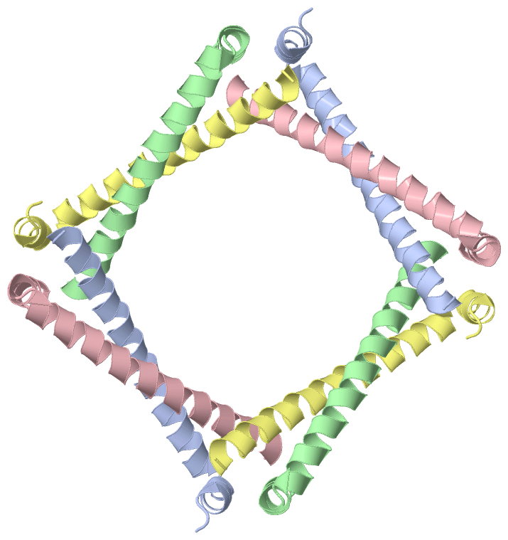
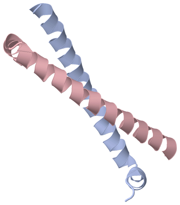
Chain A from PDB  Type:PROTEIN  Length:50
 aligned with SHDAG_HDVAM | P25989 from UniProtKB/Swiss-Prot  Length:195

    Alignment length:50
                                    21        31        41        51        61
           SHDAG_HDVAM   12 GREDILEQWVSGRKKLEELERDLRKLKKKIKKLEEDNPWLGNIKGIIGKK 61
               SCOP domains d1a92a_ A:                                         SCOP domains
               CATH domains 1a92A00 A:12-61                                    CATH domains
               Pfam domains -------------------------------------------------- Pfam domains
         Sec.struct. author .hhhhhhhhhhhhhhhhhhhhhhhhhhhhhhhhhhh.hhhhhhhhhh... Sec.struct. author
                 SAPs(SNPs) -------------------------------------------------- SAPs(SNPs)
                    PROSITE -------------------------------------------------- PROSITE
                 Transcript -------------------------------------------------- Transcript
                  1a92 A 12 GREDILEQWVSGRKKLEELERDLRKLKKKIKKLEEDNPWLGNIKGIIGKY 61
                                    21        31        41        51        61

Chain B from PDB  Type:PROTEIN  Length:50
 aligned with SHDAG_HDVAM | P25989 from UniProtKB/Swiss-Prot  Length:195

    Alignment length:50
                                    21        31        41        51        61
           SHDAG_HDVAM   12 GREDILEQWVSGRKKLEELERDLRKLKKKIKKLEEDNPWLGNIKGIIGKK 61
               SCOP domains d1a92b_ B:                                         SCOP domains
               CATH domains 1a92B00 B:12-61                                    CATH domains
               Pfam domains -------------------------------------------------- Pfam domains
         Sec.struct. author .hhhhhhhhhhhhhhhhhhhhhhhhhhhhhhhhhhh.hhhhhhhhhh... Sec.struct. author
                 SAPs(SNPs) -------------------------------------------------- SAPs(SNPs)
                    PROSITE -------------------------------------------------- PROSITE
                 Transcript -------------------------------------------------- Transcript
                  1a92 B 12 GREDILEQWVSGRKKLEELERDLRKLKKKIKKLEEDNPWLGNIKGIIGKY 61
                                    21        31        41        51        61

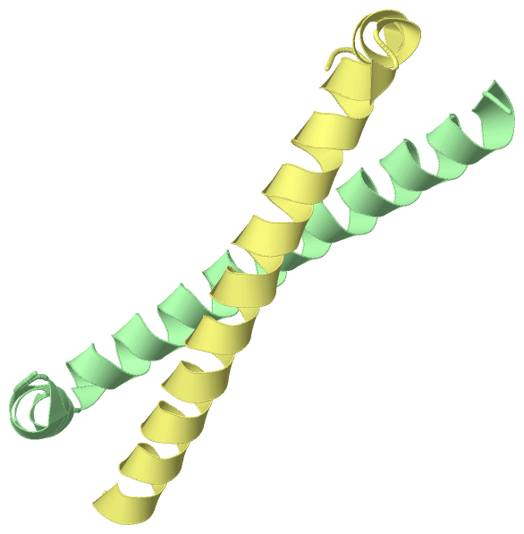
Chain C from PDB  Type:PROTEIN  Length:50
 aligned with SHDAG_HDVAM | P25989 from UniProtKB/Swiss-Prot  Length:195

    Alignment length:50
                                    21        31        41        51        61
           SHDAG_HDVAM   12 GREDILEQWVSGRKKLEELERDLRKLKKKIKKLEEDNPWLGNIKGIIGKK 61
               SCOP domains d1a92c_ C:                                         SCOP domains
               CATH domains 1a92C00 C:12-61                                    CATH domains
               Pfam domains -------------------------------------------------- Pfam domains
         Sec.struct. author .hhhhhhhhhhhhhhhhhhhhhhhhhhhhhhhhhhh.hhhhhhhhhh... Sec.struct. author
                 SAPs(SNPs) -------------------------------------------------- SAPs(SNPs)
                    PROSITE -------------------------------------------------- PROSITE
                 Transcript -------------------------------------------------- Transcript
                  1a92 C 12 GREDILEQWVSGRKKLEELERDLRKLKKKIKKLEEDNPWLGNIKGIIGKY 61
                                    21        31        41        51        61

Chain D from PDB  Type:PROTEIN  Length:49
 aligned with SHDAG_HDVAM | P25989 from UniProtKB/Swiss-Prot  Length:195

    Alignment length:49
                                    21        31        41        51         
           SHDAG_HDVAM   12 GREDILEQWVSGRKKLEELERDLRKLKKKIKKLEEDNPWLGNIKGIIGK 60
               SCOP domains d1a92d_ D:                                        SCOP domains
               CATH domains 1a92D00 D:12-60                                   CATH domains
               Pfam domains ------------------------------------------------- Pfam domains
         Sec.struct. author .hhhhhhhhhhhhhhhhhhhhhhhhhhhhhhhhhhh.hhhhhhhhh... Sec.struct. author
                 SAPs(SNPs) ------------------------------------------------- SAPs(SNPs)
                    PROSITE ------------------------------------------------- PROSITE
                 Transcript ------------------------------------------------- Transcript
                  1a92 D 12 GREDILEQWVSGRKKLEELERDLRKLKKKIKKLEEDNPWLGNIKGIIGK 60
                                    21        31        41        51         

   Legend:   → Mismatch (orange background)
  - → Gap (green background, '-', border residues have a numbering label)
    → Modified Residue (blue background, lower-case, 'x' indicates undefined single-letter code, labelled with number + name)
  x → Chemical Group (purple background, 'x', labelled with number + name, e.g. ACE or NH2)
  extra numbering lines below/above indicate numbering irregularities and modified residue names etc., number ends below/above '|'

 Classification and Annotation

(-) SCOP Domains  (1, 4)

Asymmetric Unit

(-) CATH Domains  (1, 4)

Asymmetric Unit

(-) Pfam Domains  (0, 0)

(no "Pfam Domain" information available for 1A92)

(-) Gene Ontology  (6, 6)

Asymmetric Unit(hide GO term definitions)
Chain A,B,C,D   (SHDAG_HDVAM | P25989)
molecular function
    GO:0003723    RNA binding    Interacting selectively and non-covalently with an RNA molecule or a portion thereof.
biological process
    GO:0046718    viral entry into host cell    The process that occurs after viral attachment by which a virus, or viral nucleic acid, breaches the plasma membrane or cell envelope and enters the host cell. The process ends when the viral nucleic acid is released into the host cell cytoplasm.
    GO:0075732    viral penetration into host nucleus    The crossing by the virus of the host nuclear membrane, either as naked viral genome or for small viruses as an intact capsid.
    GO:0016032    viral process    A multi-organism process in which a virus is a participant. The other participant is the host. Includes infection of a host cell, replication of the viral genome, and assembly of progeny virus particles. In some cases the viral genetic material may integrate into the host genome and only subsequently, under particular circumstances, 'complete' its life cycle.
cellular component
    GO:0042025    host cell nucleus    A membrane-bounded organelle as it is found in the host cell in which chromosomes are housed and replicated. The host is defined as the larger of the organisms involved in a symbiotic interaction.
    GO:0019012    virion    The complete fully infectious extracellular virus particle.

 Visualization

(-) Interactive Views

Asymmetric Unit
  Complete Structure
    Jena3D(integrated viewing of ligand, site, SAP, PROSITE, SCOP information)
    WebMol | AstexViewer[tm]@PDBe
(Java Applets, require no local installation except for Java; loading may be slow)
    STRAP
(Java WebStart application, automatic local installation, requires Java; full application with system access!)
    RasMol
(require local installation)
    Molscript (VRML)
(requires installation of a VRML viewer; select preferred view via VRML and generate a mono or stereo PDF format file)
 
  Ligands, Modified Residues, Ions
(no "Ligands, Modified Residues, Ions" information available for 1a92)
 
  Sites
(no "Sites" information available for 1a92)
 
  Cis Peptide Bonds
(no "Cis Peptide Bonds" information available for 1a92)
 
Biological Units
  Complete Structure
    Biological Unit 1  [ Jena3D ]
    Biological Unit 2  [ Jena3D ]
    Biological Unit 3  [ Jena3D ]

(-) Still Images

Jmol
  protein: cartoon or spacefill or dots and stick; nucleic acid: cartoon and stick; ligands: spacefill; active site: stick
Molscript
  protein, nucleic acid: cartoon; ligands: spacefill; active site: ball and stick

 Databases and Analysis Tools

(-) Databases

Access by PDB/NDB ID
  1a92
    Family and Domain InformationProDom | SYSTERS
    General Structural InformationGlycoscienceDB | MMDB | NDB | OCA | PDB | PDBe | PDBj | PDBsum | PDBWiki | PQS | PROTEOPEDIA
    Orientation in MembranesOPM
    Protein SurfaceSURFACE
    Secondary StructureDSSP (structure derived) | HSSP (homology derived)
    Structural GenomicsGeneCensus
    Structural NeighboursCE | VAST
    Structure ClassificationCATH | Dali | SCOP
    Validation and Original DataBMRB Data View | BMRB Restraints Grid | EDS | PROCHECK | RECOORD | WHAT_CHECK
 
Access by UniProt ID/Accession number
  SHDAG_HDVAM | P25989
    Comparative Protein Structure ModelsModBase
    Genomic InformationEnsembl
    Protein-protein InteractionDIP
    Sequence, Family and Domain InformationInterPro | Pfam | SMART | UniProtKB/SwissProt
 
Access by Enzyme Classificator   (EC Number)
  (no 'Enzyme Classificator' available)
    General Enzyme InformationBRENDA | EC-PDB | Enzyme | IntEnz
    PathwayKEGG | MetaCyc
 
Access by Disease Identifier   (MIM ID)
  (no 'MIM ID' available)
    Disease InformationOMIM
 
Access by GenAge ID
  (no 'GenAge ID' available)
    Age Related InformationGenAge

(-) Analysis Tools

Access by PDB/NDB ID
    Domain InformationXDom
    Interatomic Contacts of Structural UnitsCSU
    Ligand-protein ContactsLPC
    Protein CavitiescastP
    Sequence and Secondary StructurePDBCartoon
    Structure AlignmentSTRAP(Java WebStart application, automatic local installation, requires Java; full application with system access!)
    Structure and Sequence BrowserSTING
 
Access by UniProt ID/Accession number
  SHDAG_HDVAM | P25989
    Protein Disorder PredictionDisEMBL | FoldIndex | GLOBPLOT (for more information see DisProt)

 Related Entries

(-) Entries Sharing at Least One Protein Chain (UniProt ID)

UniProtKB/Swiss-Prot
        SHDAG_HDVAM | P259891by0

(-) Related Entries Specified in the PDB File

(no "Related Entries Specified in the PDB File" available for 1A92)