|
|
|
|
 Description
|
|
Compounds
|
||||||||||||||||||||||||||||||||||||||||||||||||||||||||||||||||||||||||||||||||||||||||||
Chains, Units
Summary Information (see also Sequences/Alignments below) |
Ligands, Modified Residues, Ions (0, 0)| (no "Ligand,Modified Residues,Ions" information available for 1A7O) |
Sites (0, 0)| (no "Site" information available for 1A7O) |
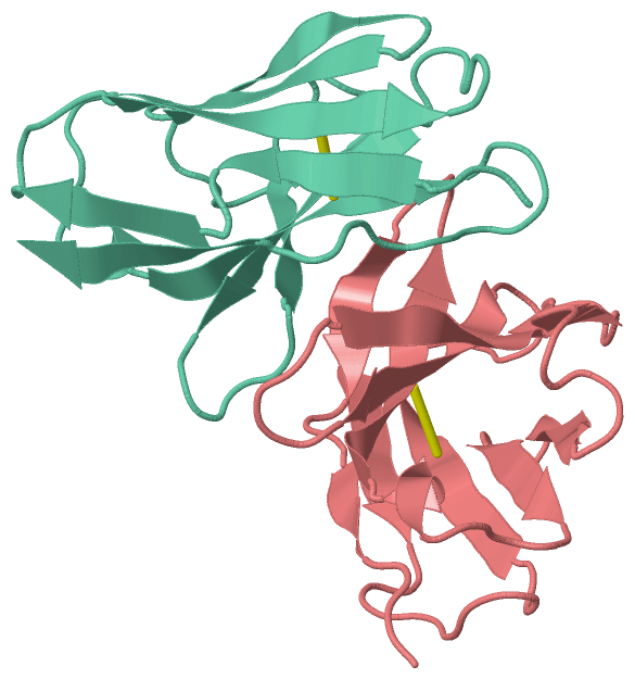
SS Bonds (2, 2)
Asymmetric/Biological Unit
|
||||||||||||
Cis Peptide Bonds (1, 1)
Asymmetric/Biological Unit
|
||||||||
SAPs(SNPs)/Variants (0, 0)| (no "SAP(SNP)/Variant" information available for 1A7O) |
PROSITE Motifs (0, 0)| (no "PROSITE Motif" information available for 1A7O) |
Exons (0, 0)| (no "Exon" information available for 1A7O) |
Sequences/Alignments
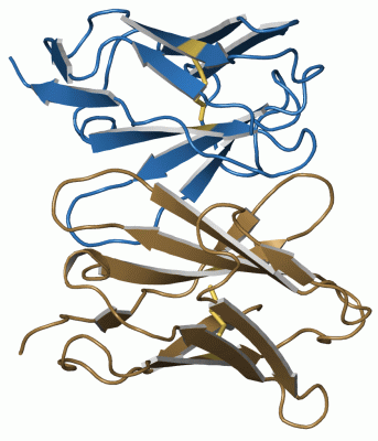
Asymmetric/Biological UnitChain H from PDB Type:PROTEIN Length:116 aligned with HVM44_MOUSE | P01820 from UniProtKB/Swiss-Prot Length:115 Alignment length:116 115 29 39 49 59 69 79 89 99 109 | - - HVM44_MOUSE 20 QVQLKESGPGLVAPSQSLSITCTVSGFSLTGYGVNWVRQPPGKGLEWLGMIWGDGSTDYNSALKSRLSISKDNSKSQVFLKMNSLQTDDTARYYCA-------------------- - SCOP domains d1a7oh_ H: Immunoglobulin heavy chain variable domain, VH SCOP domains CATH domains 1a7oH00 H:201-316 Immunoglobulins CATH domains Pfam domains -------------------------------------------------------------------------------------------------------------------- Pfam domains SAPs(SNPs) -------------------------------------------------------------------------------------------------------------------- SAPs(SNPs) PROSITE -------------------------------------------------------------------------------------------------------------------- PROSITE Transcript -------------------------------------------------------------------------------------------------------------------- Transcript 1a7o H 201 QVQLQESGPGLVAPSQSLSITCTVSGFSLTGYGVNWVRQPPGKGLEWLGMIWGDGNTDYNSALKSRLSISKDNSKSQVFLKMNSLHTDDTARYYCARERDYRLDYWGQGTTVTVSS 316 210 220 230 240 250 260 270 280 290 300 310 Chain L from PDB Type:PROTEIN Length:106 aligned with KV5A3_MOUSE | P01635 from UniProtKB/Swiss-Prot Length:115 Alignment length:106 115 30 40 50 60 70 80 90 100 110 | - KV5A3_MOUSE 21 DIQMTQSPASLSASVGETVTITCRASGNIHNYLAWYQQKQGKSPQLLVYNAKTLADGVPSRFSGSGSGTQYSLKINSLQPEDFGSYYCQHFWSTP----------- - SCOP domains d1a7ol_ L: Immunoglobulin light chain kappa variable domain, VL-kappa SCOP domains CATH domains 1a7oL00 L:1-107 Immunoglobulins CATH domains Pfam domains ---------------------------------------------------------------------------------------------------------- Pfam domains SAPs(SNPs) ---------------------------------------------------------------------------------------------------------- SAPs(SNPs) PROSITE ---------------------------------------------------------------------------------------------------------- PROSITE Transcript ---------------------------------------------------------------------------------------------------------- Transcript 1a7o L 1 DIVLTQSPASLSASVGETVTITCRASGNIHNYLAWYQQKQGKSPQLLVYYTTTLADGVPSRFSGSGSGTQYSLKINSLQPDDFGSYYCQHFWSTPTFGGGTKLEIK 107 10 20 30 40 50 60 70 80 90 || 101 95| 97
|
||||||||||||||||||||
SCOP Domains (2, 2)
Asymmetric/Biological Unit
 |
CATH Domains (1, 2)
Asymmetric/Biological Unit
|
Pfam Domains (0, 0)| (no "Pfam Domain" information available for 1A7O) |
Gene Ontology (1, 2)|
Asymmetric/Biological Unit(hide GO term definitions) Chain H (HVM44_MOUSE | P01820)
Chain L (KV5A3_MOUSE | P01635)
|
||||||||||||||||||||||||
Interactive Views
|
|||||||||||||||||||||||||||||||||||||||||||||||||||||||||||||||||||||||||||||||||||||||||||||||||||||||||||||||||||||
Still Images
|
||||||||||||||||
Databases
|
||||||||||||||||||||||||||||||||||||||||||||||||||||||||||||||||||||||||||||||||||||||||||||||||||||||||||||||||||||||||||||||||||||||||||||||||||||||||||||||||||||||||||||||||||||||||||
Analysis Tools
|
||||||||||||||||||||||||||||||||||||||||||||||||||||||||||||||||||||||||
Entries Sharing at Least One Protein Chain (UniProt ID)
Related Entries Specified in the PDB File
|
|